MoleMax Range
MoleMax HD
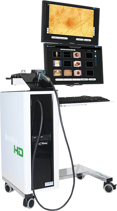
MoleMax HD is a modular digital dermoscopy system designed for use in dermatology and general practice. It supports lesion scoring, trending and monitoring, and patient reporting.
MoleMax HD is a modular digital dermoscopy system designed for use in dermatology and general practice. It supports lesion scoring, trending and monitoring, and patient reporting.
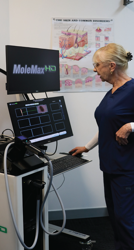
Since 1997, MoleMax has been a pioneer in digital dermoscopy, helping dermatologists and skin cancer clinics see more, detect earlier, and care better.
Developed in collaboration with leading clinicians at the University of Vienna, our systems are now used in over 60 countries, with more than 3,000 installations worldwide. From the high-definition MoleMax HD PRO to the compact MoleMax Lite, every product is designed alongside the doctors who use it.
Australian and Austrian-engineered, MoleMax delivers clinically proven imaging with no subscription fees, no cloud dependencies, and full local control of patient data. Our hardware is built to last; our software is built to work the way clinicians work.
A complete skin imaging platform. Built with clinicians, for clinicians.
Improves productivity with quick, simple follow-up and trending, supported by an extensive skin reference library, with no subscriptions or hidden costs.
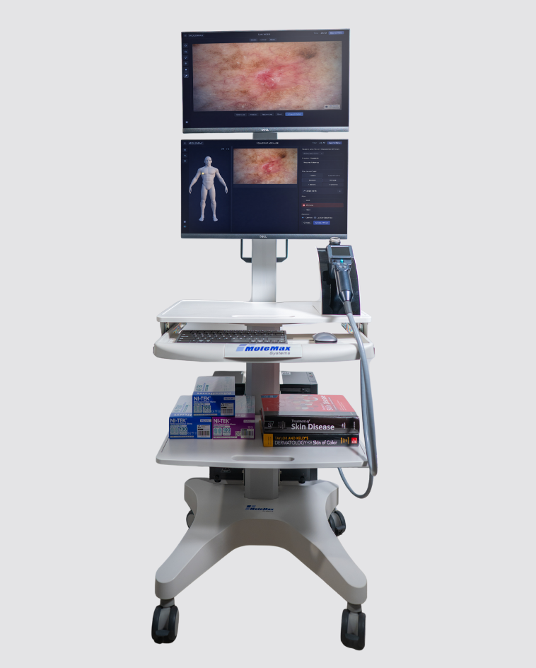
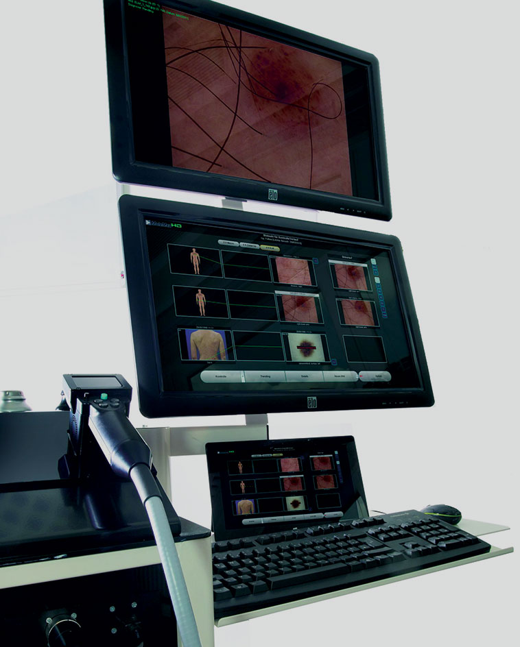
Every camera, adapter, and lens in the MoleMax HD system is designed to capture skin with clarity, consistency, and the level of detail clinicians rely on to make confident decisions.
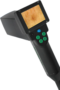
This camera is only available with the MoleMax HD. It uses Sony technology to capture the maximum image resolution available with the HD image
This camera is only available with the MoleMax HD. It uses Sony technology to capture the maximum image resolution available with the HD image
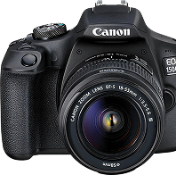
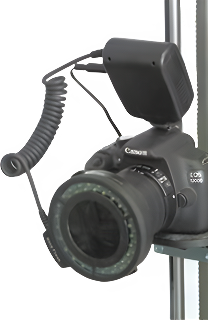
A Canon SLR is available with the MoleMax HD, Automatic Total Body Mapping Stand and PhotoMAX PRO systems...
A Canon SLR is available with the MoleMax HD, Automatic Total Body Mapping Stand and PhotoMAX PRO systems. It connects to the system via USB and provides a live image on screen before capture. This camera may also be mounted onto our body mapping stand. The high resolution digital camera can also be connected to your dermatoscope for live imaging with your MoleMax.

Interchangeable adapters that may be used with DermDOC and MoleMax Macro Cameras. From left to right: Non-P...
Interchangeable adapters that may be used with DermDOC and MoleMax Macro Cameras. From left to right: Non-Polarised 20, 30, 40x adapter (optional), Non-Polarised 60, 80, 100x adapter (optional), Polarised 20, 30, 40x adapter (standard), 60, 80, 100x Polarised adapter (optional).
Digital Epiluminescence Microscopy (DELM) using polarized light with options for wet and dry dermoscopic imaging.Even an experienced dermatologist finds it difficult to distinguish between a benign and a malignant mole in the early stages.
Digital Epiluminescence Microscopy (DELM) using polarized light with options for wet and dry dermoscopic imaging. Even an experienced dermatologist finds it difficult to distinguish between a benign and a malignant mole in the early stages. A more detailed and precise diagnosis requires skin surface microscopy. The method of epiluminescence microscopy opens up a new view of morphological features, colours and patterns not seen in an examination with traditional skin surface microscopy. The University of Vienna Medical School in Austria has pioneered the introduction of this advanced method. The MoleMax developers advanced this technology even further by developing the only fully hygienically dermoscopy method available, using cross polarised light.
From diagnosis libraries to follow-up trending, MoleMax HD software brings documentation,
comparison, and reporting into one efficient workflow, with data that stays under your control.

The MoleMax and SkinDOC systems runs perfectly within most clinic networks. Data can be stored and backed up on a clinics’ server. Where a spare server is not available, the MoleMax is equipped with...
The MoleMax and SkinDOC systems runs perfectly within most clinic networks. Data can be stored and backed up on a clinics’ server. Where a spare server is not available, the MoleMax is equipped with ports for external backup. Patient images and files may also be exported from the MoleMax system into practice management software.

The All Skin module is a diagnosis library
filled with images and descriptions which
give the user the ability to browse and
search for certain skin conditions. The skin
conditions vary from melanoma to actini...
The All Skin module is a diagnosis library filled with images and descriptions which give the user the ability to browse and search for certain skin conditions. The skin conditions vary from melanoma to actinic keratosis, basal cell carcinomas and many more. The skin library features hundreds of them, including those not related to skin cancer.

The Histopathology module is primarily
used to import Histo images into the
patient database. Histo images may be
imported from any storage media
connected to the MoleMax or SkinDOC s...
The Histopathology module is primarily used to import Histo images into the patient database. Histo images may be imported from any storage media connected to the MoleMax or SkinDOC systems including the local drive, CD/DVD drive, a USB or flash drive, external storage drive or a network location.

A quick comparison in large scale is one of
the most important features Doctors look
for in Skin Imaging software. A follow-up
image may be taken with the previous
image in view on one side of the screen....
A quick comparison in large scale is one of the most important features Doctors look for in Skin Imaging software. A follow-up image may be taken with the previous image in view on one side of the screen. Alternatively, an outline may be generated of the original image to allow for easier follow-up. Images over time may also be compared side by side or over each other to easily assist in recognising lesion alterations.

The TrichoScan software allows you to
automatically measure and calculate
biological parameters of hair growth.
Images taken with the MoleMAX or com...
The TrichoScan software allows you to automatically measure and calculate biological parameters of hair growth. Images taken with the MoleMAX or compatible SLR camera are analysed for changes. The TrichoScan module is an ideal tool to monitor hair loss and treatment response. Some of the parameters that the TrichoScan module assists include hair density, diameter, growth rate and Anagen/Telegen ratio.
*An optional extra. Additional charges apply.
When the user requires more than a
manual scoring method, the Automatic
Scoring Module can assist further. This
module is used for measurement of mol...
When the user requires more than a manual scoring method, the Automatic Scoring Module can assist further. This module is used for measurement of moles and pigmented skin lesions. It is a suitable second option, calculating the risk level using the world wide accepted ABCD rule. The software module automatically calculates each parameter of the ABCD rule and provides a score and risk graph.
*An optional extra. Additional charges apply.
The Thumbnail follow up feature is a new
addition created by Doctor and User
feedback. This popular new module
allows follow ups to be taken from a
different view which combines onscreen...
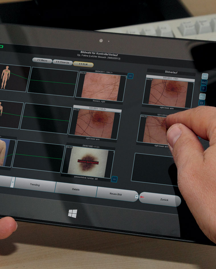
The Thumbnail follow up feature is a new addition created by Doctor and User feedback. This popular new module allows follow ups to be taken from a different view which combines onscreen the micro image, macro image and dummy location at the same time to allow for quick and easy follow up sessions. Images taken may be scored as either high, medium or low risk and these can be sorted on screen for simultaneous view based on the risk. All micro and macro images taken may also have all their follow-up images appear side by side on the screen to easily identify the changes over time via the trending approach.

This module contains a histopathology
database of diagnosed pigmented lesions
taken on the MoleMax system. These
diagnosed images can be used for
comparison and scoring purposes. This...
This module contains a histopathology database of diagnosed pigmented lesions taken on the MoleMax system. These diagnosed images can be used for comparison and scoring purposes. This module also includes image analysis functions which will provide the user with automated calculations for various parameters such as diameter, circumference, and area measurements. The module can also auto-measure and compare any two lesions taken over time to analyse pattern and size changes. The aid-to-diagnose module has in-built scoring tools which guide the user to assess risk of a lesion with a score.

Two similar follow-up images of a relevant
body site for one patient will be compared
as it can be difficult to identify changed or
new mole. The mole mapping module a...
Two similar follow-up images of a relevant body site for one patient will be compared as it can be difficult to identify changed or new mole. The mole mapping module automatically detects nevi in the first and in the follow-up image. Additionally the software extracts features such as size, shape, and brightness that are relevant to each lesion and these features are compared for all mapped nevi. Extracted moles are shown in full resolution giving a detailed image of the nevus.
*An optional extra. Additional charges apply.
Macro Trending allows for Micro and
Histopathology images to be taken over
time with comments. A complete patient
history can be kept in chronological order
every image taken and every comment a...
Macro Trending allows for Micro and Histopathology images to be taken over time with comments. A complete patient history can be kept in chronological order every image taken and every comment added to the patient file.

Our software is specially designed to
operate in a multiuser environment to
maintain audit paths and patient data
security. The Administrator may tailor
access rights for each user, to limit read,...
Our software is specially designed to operate in a multiuser environment to maintain audit paths and patient data security. The Administrator may tailor access rights for each user, to limit read, write and delete access.

This function allows the user to make a
complete photographic documentation of
the entire body based on 33 present
segments (quick examination -10
segments). This software allows importin...
This function allows the user to make a complete photographic documentation of the entire body based on 33 present segments (quick examination -10 segments). This software allows importing of images from other digital sources such as digital cameras.
MoleMax Systems
Our Practice Management Link will improve the workflow, cost efficiency and time management of your practice, linking the MoleMax software to your Practice Management Software and integrate with your patient notes.
 Before You Book
MoleMax offers a comprehensive solution for skin imaging, combining total body photography with detailed lesion tracking and documentation, all streamlined into a single, efficient workflow.
MoleMax Systems, a division of Macquarie Medical Systems, specialises in the development and production of dermatoscopic hardware and software, including dermatoscopes and the MoleMax Dermoscopy Software. These integrated solutions are designed for early clinical imaging sessions, analysis and comparisons of skin cancer, providing dermatologists with advanced tools for high-resolution imaging and analysis of skin lesions to improve patient outcomes.
Subscribe To The MoleMax Newsletter