MoleMax HD Pro Total Body Mapping System
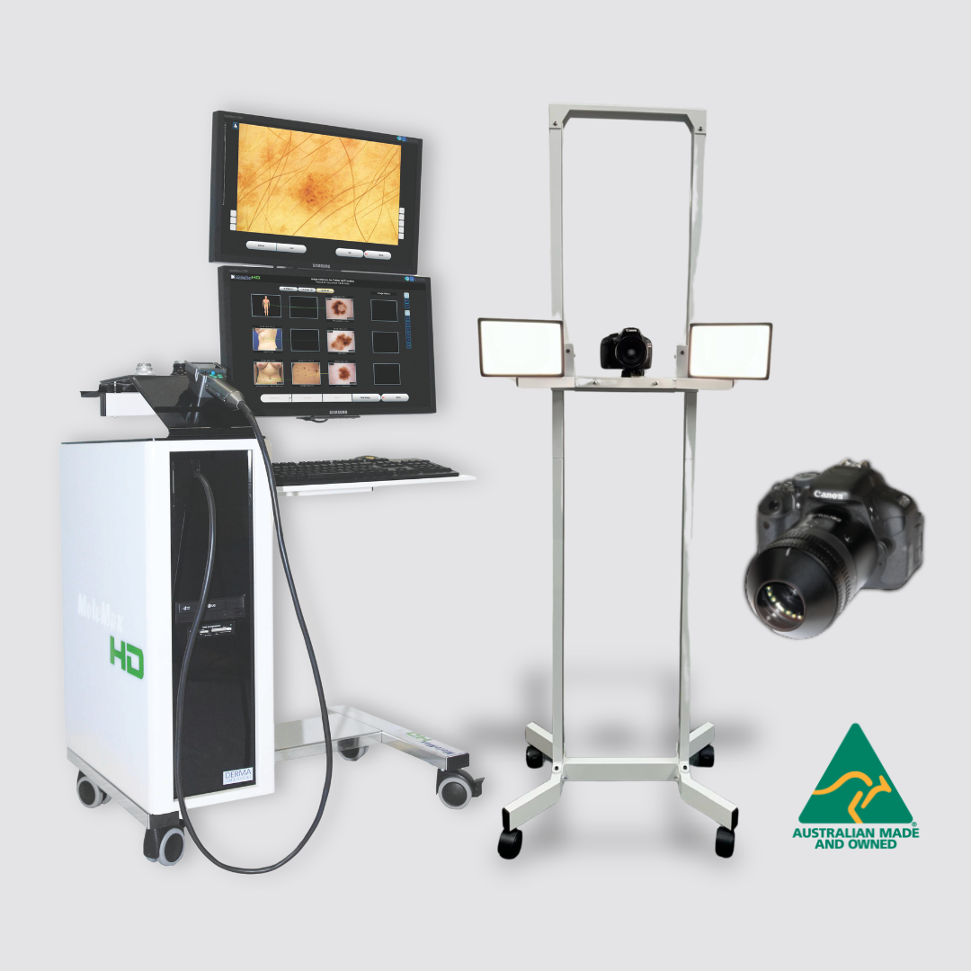
The MoleMax HD Pro is our proven legacy system for clinics seeking a complete Total Body Mapping and digital dermoscopy solution. It brings full-body photography, detailed lesion imaging and follow-up comparisons together in one streamlined platform.
Designed for busy skin cancer practices, the HD Pro helps clinicians create a comprehensive visual record of each patient’s skin. This then enables capture of baseline and follow-up images for side-by-side comparison by clinicians.

Key Benefits
Molemax HD Pro Total Body Mapping System
- Complete Total Body Mapping and digital dermoscopy in one system
- High-quality dermoscopic, macro and clinical imaging
- Configurable magnification up to 100x, with 20x, 30x, 40x, 60x, 80x and 100x options
- Efficient baseline documentation and follow-up comparisons
- Side-by-side monitoring of lesions over time
- Streamlined patient management, reporting and image storage
- Multiple screens and touchscreen operation for an efficient workflow
- Mobile trolley design suitable for dedicated imaging rooms
- No subscription fees or ongoing software costs
- Supported locally by an experienced Australian service and training team
Built on more than 25 years of MoleMax skin-imaging expertise, the HD Pro is a trusted, comprehensive solution for clinics looking to establish or expand their Total Body Mapping services.
Hardware & Software Features
MoleMax Systems

High Definition Video Camera
Read More
This camera is only available with the MoleMax HD. It uses Sony technology to capture the maximum image resolution available with the HD image quality. The camera has an integrated LCD screen to streamline the screening process. Variable adapters are available for zoom functions up to 100x. The camera is easily controlled via the buttons on the back allowing for zoom, light and all mouse controls. The camera allows for 3 types of images:
- Macro mode: Macro and close up images can be taken with a live video preview on screen. There is a light source on the camera as well as mouse controls on the back of the handle to provide quick, effective use.
- Micro mode (NO OIL): With a simple click of an adapter, you are able to view polarised images of your patients lesions with magnifications of 20x, 30x or 40x zoom. No immersion fluids are required as the adapter uses patented polarised light technology to go beneath the skin to capture an image. Also available is our high magnification adapter with 60x, 80x or 100x zoom.
- Micro mode (OIL/FLUIDS): If you prefer to use oil, we give you that option also. Available is a standard non-polarised adapter (20x, 30x 40x zoom) or a high magnification non-polarised adapter (60x, 80x, 100x zoom).

High Res Digital SLR Camera & Adapter
Read More
A Canon SLR is available with the MoleMax HD, Automatic Total Body Mapping Stand and PhotoMAX PRO systems. It connects to the system via USB and provides a live image on screen before capture. This camera may also be mounted onto our body mapping stand. The high resolution digital camera can also be connected to your dermatoscope for live imaging with your MoleMax.

MoleMax HD Adapters
Read More
Interchangeable adapters that may be used with DermDOC and MoleMax Macro Cameras. From left to right: Non-Polarised 20, 30, 40x adapter (optional), Non-Polarised 60, 80, 100x adapter (optional), Polarised 20, 30, 40x adapter (standard), 60, 80, 100x Polarised adapter (optional).

DELM Method Advanced by MoleMax Technology
Read More
Digital Epiluminescence Microscopy (DELM) using polarized light with options for wet and dry dermoscopic imaging. Even an experienced dermatologist finds it difficult to distinguish between a benign and a malignant mole in the early stages. A more detailed and precise diagnosis requires skin surface microscopy. The method of epiluminescence microscopy opens up a new view of morphological features, colours and patterns not seen in an examination with traditional skin surface microscopy. The University of Vienna Medical School in Austria has pioneered the introduction of this advanced method. The MoleMax developers advanced this technology even further by developing the only fully hygienically dermoscopy method available, using cross polarised light.

Data Back Up & Export
Read More
The MoleMax and SkinDOC systems runs perfectly within most clinic networks. Data can be stored and backed up on a clinics’ server. Where a spare server is not available, the MoleMax is equipped with ports for external backup. Patient images and files may also be exported from the MoleMax system into practice management software.

All Skin Library
Read More
The All Skin module is a diagnosis library filled with images and descriptions which give the user the ability to browse and search for certain skin conditions. The skin conditions vary from melanoma to actinic keratosis, basal cell carcinomas and many more. The skin library features hundreds of them, including those not related to skin cancer.

Histopathology
Read More
The Histopathology module is primarily used to import Histo images into the patient database. Histo images may be imported from any storage media connected to the MoleMax or SkinDOC systems including the local drive, CD/DVD drive, a USB or flash drive, external storage drive or a network location.

Real Time/Overlay Follow Up
Read More
A quick comparison in large scale is one of the most important features Doctors look for in Skin Imaging software. A follow-up image may be taken with the previous image in view on one side of the screen. Alternatively, an outline may be generated of the original image to allow for easier follow-up. Images over time may also be compared side by side or over each other to easily assist in recognising lesion alterations.

Thumbnail Monitoring
Read More
The Thumbnail follow up feature is a new addition created by Doctor and User feedback. This popular new module allows follow ups to be taken from a different view which combines onscreen the micro image, macro image and dummy location at the same time to allow for quick and easy follow up sessions. Images taken may be scored as either high, medium or low risk and these can be sorted on screen for simultaneous view based on the risk. All micro and macro images taken may also have all their follow-up images appear side by side on the screen to easily identify the changes over time via the trending approach.

Total Body Mapping
Read More
This function allows the user to make a complete photographic documentation of the entire body based on 33 present segments (quick examination -10 segments). This software allows importing of images from other digital sources such as digital cameras.

Trending and Monitoring
Read More
Macro Trending allows for Micro and Histopathology images to be taken over time with comments. A complete patient history can be kept in chronological order every image taken and every comment added to the patient file.

Multi-User Environment
Read More
Our software is specially designed to operate in a multiuser environment to maintain audit paths and patient data security. The Administrator may tailor access rights for each user, to limit read, write and delete access.

What Our Clients Say
MoleMax Systems
Seamless Practice Management Software Integration with MoleMax PDF Reports
Streamline your clinical workflow with MoleMax’s advanced PDF reporting capabilities. Generate comprehensive, high-quality reports featuring dermoscopic & clinical images along with findings, ready to be easily saved or uploaded to any Practice Management Software.
Enhance practice efficiency, simplify clinical documentation and maintain organised patient records by keeping MoleMax reports alongside existing patient notes—all supporting a more connected, efficient and streamlined patient management experience.
MoleMax HD Pro Total Body Mapping System
Advanced Total Body Mapping for Complete Skin Documentation and Monitoring
The MoleMax HD Pro is our proven legacy system for clinics seeking a complete Total Body Mapping and digital dermoscopy solution. It brings full-body photography, detailed lesion imaging and follow-up comparisons together in one streamlined platform.
Designed for busy skin cancer practices, the HD Pro helps clinicians create a comprehensive visual record of each patient’s skin. This then enables capture of baseline and follow-up images for side-by-side comparison by clinicians.
Key Benefits
Molemax HD Pro Total Body Mapping System
- Complete Total Body Mapping and digital dermoscopy in one system
- High-quality dermoscopic, macro and clinical imaging
- Configurable magnification up to 100x, with 20x, 30x, 40x, 60x, 80x and 100x options
- Efficient baseline documentation and follow-up comparisons
- Side-by-side monitoring of lesions over time
- Streamlined patient management, reporting and image storage
- Multiple screens and touchscreen operation for an efficient workflow
- Mobile trolley design suitable for dedicated imaging rooms
- No subscription fees or ongoing software costs
- Supported locally by an experienced Australian service and training team
Built on more than 25 years of MoleMax skin-imaging expertise, the HD Pro is a trusted, comprehensive solution for clinics looking to establish or expand their Total Body Mapping services.
Hardware & Software Features
MoleMax Systems
High Definition Video Camera
Read More
This camera is only available with the MoleMax HD. It uses Sony technology to capture the maximum image resolution available with the HD image quality. The camera has an integrated LCD screen to streamline the screening process. Variable adapters are available for zoom functions up to 100x. The camera is easily controlled via the buttons on the back allowing for zoom, light and all mouse controls. The camera allows for 3 types of images:
- Macro mode: Macro and close up images can be taken with a live video preview on screen. There is a light source on the camera as well as mouse controls on the back of the handle to provide quick, effective use.
- Micro mode (NO OIL): With a simple click of an adapter, you are able to view polarised images of your patients lesions with magnifications of 20x, 30x or 40x zoom. No immersion fluids are required as the adapter uses patented polarised light technology to go beneath the skin to capture an image. Also available is our high magnification adapter with 60x, 80x or 100x zoom.
- Micro mode (OIL/FLUIDS): If you prefer to use oil, we give you that option also. Available is a standard non-polarised adapter (20x, 30x 40x zoom) or a high magnification non-polarised adapter (60x, 80x, 100x zoom).
High Res Digital SLR Camera & Adapter
Read More
A Canon SLR is available with the MoleMax HD, Automatic Total Body Mapping Stand and PhotoMAX PRO systems. It connects to the system via USB and provides a live image on screen before capture. This camera may also be mounted onto our body mapping stand. The high resolution digital camera can also be connected to your dermatoscope for live imaging with your MoleMax.
MoleMax HD Adapters
Read More
Interchangeable adapters that may be used with DermDOC and MoleMax Macro Cameras. From left to right: Non-Polarised 20, 30, 40x adapter (optional), Non-Polarised 60, 80, 100x adapter (optional), Polarised 20, 30, 40x adapter (standard), 60, 80, 100x Polarised adapter (optional).
DELM Method Advanced by MoleMax Technology
Read More
Digital Epiluminescence Microscopy (DELM) using polarized light with options for wet and dry dermoscopic imaging. Even an experienced dermatologist finds it difficult to distinguish between a benign and a malignant mole in the early stages. A more detailed and precise diagnosis requires skin surface microscopy. The method of epiluminescence microscopy opens up a new view of morphological features, colours and patterns not seen in an examination with traditional skin surface microscopy. The University of Vienna Medical School in Austria has pioneered the introduction of this advanced method. The MoleMax developers advanced this technology even further by developing the only fully hygienically dermoscopy method available, using cross polarised light.
Data Back Up & Export
Read More
The MoleMax and SkinDOC systems runs perfectly within most clinic networks. Data can be stored and backed up on a clinics’ server. Where a spare server is not available, the MoleMax is equipped with ports for external backup. Patient images and files may also be exported from the MoleMax system into practice management software.
All Skin Library
Read More
The All Skin module is a diagnosis library filled with images and descriptions which give the user the ability to browse and search for certain skin conditions. The skin conditions vary from melanoma to actinic keratosis, basal cell carcinomas and many more. The skin library features hundreds of them, including those not related to skin cancer.
Histopathology
Read More
The Histopathology module is primarily used to import Histo images into the patient database. Histo images may be imported from any storage media connected to the MoleMax or SkinDOC systems including the local drive, CD/DVD drive, a USB or flash drive, external storage drive or a network location.
Real Time/Overlay Follow Up
Read More
A quick comparison in large scale is one of the most important features Doctors look for in Skin Imaging software. A follow-up image may be taken with the previous image in view on one side of the screen. Alternatively, an outline may be generated of the original image to allow for easier follow-up. Images over time may also be compared side by side or over each other to easily assist in recognising lesion alterations.
Thumbnail Monitoring
Read More
The Thumbnail follow up feature is a new addition created by Doctor and User feedback. This popular new module allows follow ups to be taken from a different view which combines onscreen the micro image, macro image and dummy location at the same time to allow for quick and easy follow up sessions. Images taken may be scored as either high, medium or low risk and these can be sorted on screen for simultaneous view based on the risk. All micro and macro images taken may also have all their follow-up images appear side by side on the screen to easily identify the changes over time via the trending approach.
Total Body Mapping
Read More
This function allows the user to make a complete photographic documentation of the entire body based on 33 present segments (quick examination -10 segments). This software allows importing of images from other digital sources such as digital cameras.
Trending and Monitoring
Read More
Macro Trending allows for Micro and Histopathology images to be taken over time with comments. A complete patient history can be kept in chronological order every image taken and every comment added to the patient file.
Multi-User Environment
Read More
Our software is specially designed to operate in a multiuser environment to maintain audit paths and patient data security. The Administrator may tailor access rights for each user, to limit read, write and delete access.
What Our Clients Say
MoleMax Systems
Seamless Practice Management Software Integration with MoleMax PDF Reports
Streamline your clinical workflow with MoleMax’s advanced PDF reporting capabilities. Generate comprehensive, high-quality reports featuring dermoscopic & clinical images along with findings, ready to be easily saved or uploaded to any Practice Management Software.
Enhance practice efficiency, simplify clinical documentation and maintain organised patient records by keeping MoleMax reports alongside existing patient notes—all supporting a more connected, efficient and streamlined patient management experience.
MoleMax HD Pro Total Body Mapping System
Advanced Total Body Mapping for Complete Skin Documentation and Monitoring
The MoleMax HD Pro is our proven legacy system for clinics seeking a complete Total Body Mapping and digital dermoscopy solution. It brings full-body photography, detailed lesion imaging and follow-up comparisons together in one streamlined platform.
Designed for busy skin cancer practices, the HD Pro helps clinicians create a comprehensive visual record of each patient’s skin. This then enables capture of baseline and follow-up images for side-by-side comparison by clinicians.
Key Benefits
Molemax HD Pro Total Body Mapping System
- Complete Total Body Mapping and digital dermoscopy in one system
- High-quality dermoscopic, macro and clinical imaging
- Configurable magnification up to 100x, with 20x, 30x, 40x, 60x, 80x and 100x options
- Efficient baseline documentation and follow-up comparisons
- Side-by-side monitoring of lesions over time
- Streamlined patient management, reporting and image storage
- Multiple screens and touchscreen operation for an efficient workflow
- Mobile trolley design suitable for dedicated imaging rooms
- No subscription fees or ongoing software costs
- Supported locally by an experienced Australian service and training team
Built on more than 25 years of MoleMax skin-imaging expertise, the HD Pro is a trusted, comprehensive solution for clinics looking to establish or expand their Total Body Mapping services.
Hardware & Software Features
MoleMax Systems
High Definition Video Camera
Read More
This camera is only available with the MoleMax HD. It uses Sony technology to capture the maximum image resolution available with the HD image quality. The camera has an integrated LCD screen to streamline the screening process. Variable adapters are available for zoom functions up to 100x. The camera is easily controlled via the buttons on the back allowing for zoom, light and all mouse controls. The camera allows for 3 types of images:
- Macro mode: Macro and close up images can be taken with a live video preview on screen. There is a light source on the camera as well as mouse controls on the back of the handle to provide quick, effective use.
- Micro mode (NO OIL): With a simple click of an adapter, you are able to view polarised images of your patients lesions with magnifications of 20x, 30x or 40x zoom. No immersion fluids are required as the adapter uses patented polarised light technology to go beneath the skin to capture an image. Also available is our high magnification adapter with 60x, 80x or 100x zoom.
- Micro mode (OIL/FLUIDS): If you prefer to use oil, we give you that option also. Available is a standard non-polarised adapter (20x, 30x 40x zoom) or a high magnification non-polarised adapter (60x, 80x, 100x zoom).
High Res Digital SLR Camera & Adapter
Read More
A Canon SLR is available with the MoleMax HD, Automatic Total Body Mapping Stand and PhotoMAX PRO systems. It connects to the system via USB and provides a live image on screen before capture. This camera may also be mounted onto our body mapping stand. The high resolution digital camera can also be connected to your dermatoscope for live imaging with your MoleMax.
MoleMax HD Adapters
Read More
Interchangeable adapters that may be used with DermDOC and MoleMax Macro Cameras. From left to right: Non-Polarised 20, 30, 40x adapter (optional), Non-Polarised 60, 80, 100x adapter (optional), Polarised 20, 30, 40x adapter (standard), 60, 80, 100x Polarised adapter (optional).
DELM Method Advanced by MoleMax Technology
Read More
Digital Epiluminescence Microscopy (DELM) using polarized light with options for wet and dry dermoscopic imaging. Even an experienced dermatologist finds it difficult to distinguish between a benign and a malignant mole in the early stages. A more detailed and precise diagnosis requires skin surface microscopy. The method of epiluminescence microscopy opens up a new view of morphological features, colours and patterns not seen in an examination with traditional skin surface microscopy. The University of Vienna Medical School in Austria has pioneered the introduction of this advanced method. The MoleMax developers advanced this technology even further by developing the only fully hygienically dermoscopy method available, using cross polarised light.
Data Back Up & Export
Read More
The MoleMax and SkinDOC systems runs perfectly within most clinic networks. Data can be stored and backed up on a clinics’ server. Where a spare server is not available, the MoleMax is equipped with ports for external backup. Patient images and files may also be exported from the MoleMax system into practice management software.
All Skin Library
Read More
The All Skin module is a diagnosis library filled with images and descriptions which give the user the ability to browse and search for certain skin conditions. The skin conditions vary from melanoma to actinic keratosis, basal cell carcinomas and many more. The skin library features hundreds of them, including those not related to skin cancer.
Histopathology
Read More
The Histopathology module is primarily used to import Histo images into the patient database. Histo images may be imported from any storage media connected to the MoleMax or SkinDOC systems including the local drive, CD/DVD drive, a USB or flash drive, external storage drive or a network location.
Real Time/Overlay Follow Up
Read More
A quick comparison in large scale is one of the most important features Doctors look for in Skin Imaging software. A follow-up image may be taken with the previous image in view on one side of the screen. Alternatively, an outline may be generated of the original image to allow for easier follow-up. Images over time may also be compared side by side or over each other to easily assist in recognising lesion alterations.
Thumbnail Monitoring
Read More
The Thumbnail follow up feature is a new addition created by Doctor and User feedback. This popular new module allows follow ups to be taken from a different view which combines onscreen the micro image, macro image and dummy location at the same time to allow for quick and easy follow up sessions. Images taken may be scored as either high, medium or low risk and these can be sorted on screen for simultaneous view based on the risk. All micro and macro images taken may also have all their follow-up images appear side by side on the screen to easily identify the changes over time via the trending approach.
Total Body Mapping
Read More
This function allows the user to make a complete photographic documentation of the entire body based on 33 present segments (quick examination -10 segments). This software allows importing of images from other digital sources such as digital cameras.
Trending and Monitoring
Read More
Macro Trending allows for Micro and Histopathology images to be taken over time with comments. A complete patient history can be kept in chronological order every image taken and every comment added to the patient file.
Multi-User Environment
Read More
Our software is specially designed to operate in a multiuser environment to maintain audit paths and patient data security. The Administrator may tailor access rights for each user, to limit read, write and delete access.
What Our Clients Say
MoleMax Systems
Seamless Practice Management Software Integration with MoleMax PDF Reports
Streamline your clinical workflow with MoleMax’s advanced PDF reporting capabilities. Generate comprehensive, high-quality reports featuring dermoscopic & clinical images along with findings, ready to be easily saved or uploaded to any Practice Management Software.
Enhance practice efficiency, simplify clinical documentation and maintain organised patient records by keeping MoleMax reports alongside existing patient notes—all supporting a more connected, efficient and streamlined patient management experience.
MoleMax HD Pro Total Body Mapping System
Advanced Total Body Mapping for Complete Skin Documentation and Monitoring
The MoleMax HD Pro is our proven legacy system for clinics seeking a complete Total Body Mapping and digital dermoscopy solution. It brings full-body photography, detailed lesion imaging and follow-up comparisons together in one streamlined platform.
Designed for busy skin cancer practices, the HD Pro helps clinicians create a comprehensive visual record of each patient’s skin. This then enables capture of baseline and follow-up images for side-by-side comparison by clinicians.
Key Benefits
Molemax HD Pro Total Body Mapping System
- Complete Total Body Mapping and digital dermoscopy in one system
- High-quality dermoscopic, macro and clinical imaging
- Configurable magnification up to 100x, with 20x, 30x, 40x, 60x, 80x and 100x options
- Efficient baseline documentation and follow-up comparisons
- Side-by-side monitoring of lesions over time
- Streamlined patient management, reporting and image storage
- Multiple screens and touchscreen operation for an efficient workflow
- Mobile trolley design suitable for dedicated imaging rooms
- No subscription fees or ongoing software costs
- Supported locally by an experienced Australian service and training team
Built on more than 25 years of MoleMax skin-imaging expertise, the HD Pro is a trusted, comprehensive solution for clinics looking to establish or expand their Total Body Mapping services.
Hardware & Software Features
MoleMax Systems
High Definition Video Camera
Read More
This camera is only available with the MoleMax HD. It uses Sony technology to capture the maximum image resolution available with the HD image quality. The camera has an integrated LCD screen to streamline the screening process. Variable adapters are available for zoom functions up to 100x. The camera is easily controlled via the buttons on the back allowing for zoom, light and all mouse controls. The camera allows for 3 types of images:
- Macro mode: Macro and close up images can be taken with a live video preview on screen. There is a light source on the camera as well as mouse controls on the back of the handle to provide quick, effective use.
- Micro mode (NO OIL): With a simple click of an adapter, you are able to view polarised images of your patients lesions with magnifications of 20x, 30x or 40x zoom. No immersion fluids are required as the adapter uses patented polarised light technology to go beneath the skin to capture an image. Also available is our high magnification adapter with 60x, 80x or 100x zoom.
- Micro mode (OIL/FLUIDS): If you prefer to use oil, we give you that option also. Available is a standard non-polarised adapter (20x, 30x 40x zoom) or a high magnification non-polarised adapter (60x, 80x, 100x zoom).
High Res Digital SLR Camera & Adapter
Read More
A Canon SLR is available with the MoleMax HD, Automatic Total Body Mapping Stand and PhotoMAX PRO systems. It connects to the system via USB and provides a live image on screen before capture. This camera may also be mounted onto our body mapping stand. The high resolution digital camera can also be connected to your dermatoscope for live imaging with your MoleMax.
MoleMax HD Adapters
Read More
Interchangeable adapters that may be used with DermDOC and MoleMax Macro Cameras. From left to right: Non-Polarised 20, 30, 40x adapter (optional), Non-Polarised 60, 80, 100x adapter (optional), Polarised 20, 30, 40x adapter (standard), 60, 80, 100x Polarised adapter (optional).
DELM Method Advanced by MoleMax Technology
Read More
Digital Epiluminescence Microscopy (DELM) using polarized light with options for wet and dry dermoscopic imaging. Even an experienced dermatologist finds it difficult to distinguish between a benign and a malignant mole in the early stages. A more detailed and precise diagnosis requires skin surface microscopy. The method of epiluminescence microscopy opens up a new view of morphological features, colours and patterns not seen in an examination with traditional skin surface microscopy. The University of Vienna Medical School in Austria has pioneered the introduction of this advanced method. The MoleMax developers advanced this technology even further by developing the only fully hygienically dermoscopy method available, using cross polarised light.
Data Back Up & Export
Read More
The MoleMax and SkinDOC systems runs perfectly within most clinic networks. Data can be stored and backed up on a clinics’ server. Where a spare server is not available, the MoleMax is equipped with ports for external backup. Patient images and files may also be exported from the MoleMax system into practice management software.
All Skin Library
Read More
The All Skin module is a diagnosis library filled with images and descriptions which give the user the ability to browse and search for certain skin conditions. The skin conditions vary from melanoma to actinic keratosis, basal cell carcinomas and many more. The skin library features hundreds of them, including those not related to skin cancer.
Histopathology
Read More
The Histopathology module is primarily used to import Histo images into the patient database. Histo images may be imported from any storage media connected to the MoleMax or SkinDOC systems including the local drive, CD/DVD drive, a USB or flash drive, external storage drive or a network location.
Real Time/Overlay Follow Up
Read More
A quick comparison in large scale is one of the most important features Doctors look for in Skin Imaging software. A follow-up image may be taken with the previous image in view on one side of the screen. Alternatively, an outline may be generated of the original image to allow for easier follow-up. Images over time may also be compared side by side or over each other to easily assist in recognising lesion alterations.
Thumbnail Monitoring
Read More
The Thumbnail follow up feature is a new addition created by Doctor and User feedback. This popular new module allows follow ups to be taken from a different view which combines onscreen the micro image, macro image and dummy location at the same time to allow for quick and easy follow up sessions. Images taken may be scored as either high, medium or low risk and these can be sorted on screen for simultaneous view based on the risk. All micro and macro images taken may also have all their follow-up images appear side by side on the screen to easily identify the changes over time via the trending approach.
Total Body Mapping
Read More
This function allows the user to make a complete photographic documentation of the entire body based on 33 present segments (quick examination -10 segments). This software allows importing of images from other digital sources such as digital cameras.
Trending and Monitoring
Read More
Macro Trending allows for Micro and Histopathology images to be taken over time with comments. A complete patient history can be kept in chronological order every image taken and every comment added to the patient file.
Multi-User Environment
Read More
Our software is specially designed to operate in a multiuser environment to maintain audit paths and patient data security. The Administrator may tailor access rights for each user, to limit read, write and delete access.
What Our Clients Say
MoleMax Systems
Seamless Practice Management Software Integration with MoleMax PDF Reports
Streamline your clinical workflow with MoleMax’s advanced PDF reporting capabilities. Generate comprehensive, high-quality reports featuring dermoscopic & clinical images along with findings, ready to be easily saved or uploaded to any Practice Management Software.
Enhance practice efficiency, simplify clinical documentation and maintain organised patient records by keeping MoleMax reports alongside existing patient notes—all supporting a more connected, efficient and streamlined patient management experience.
MoleMax HD Pro Total Body Mapping System
Advanced Total Body Mapping for Complete Skin Documentation and Monitoring
The MoleMax HD Pro is our proven legacy system for clinics seeking a complete Total Body Mapping and digital dermoscopy solution. It brings full-body photography, detailed lesion imaging and follow-up comparisons together in one streamlined platform.
Designed for busy skin cancer practices, the HD Pro helps clinicians create a comprehensive visual record of each patient’s skin. This then enables capture of baseline and follow-up images for side-by-side comparison by clinicians.
Key Benefits
Molemax HD Pro Total Body Mapping System
- Complete Total Body Mapping and digital dermoscopy in one system
- High-quality dermoscopic, macro and clinical imaging
- Configurable magnification up to 100x, with 20x, 30x, 40x, 60x, 80x and 100x options
- Efficient baseline documentation and follow-up comparisons
- Side-by-side monitoring of lesions over time
- Streamlined patient management, reporting and image storage
- Multiple screens and touchscreen operation for an efficient workflow
- Mobile trolley design suitable for dedicated imaging rooms
- No subscription fees or ongoing software costs
- Supported locally by an experienced Australian service and training team
Built on more than 25 years of MoleMax skin-imaging expertise, the HD Pro is a trusted, comprehensive solution for clinics looking to establish or expand their Total Body Mapping services.
Hardware & Software Features
MoleMax Systems
High Definition Video Camera
Read More
This camera is only available with the MoleMax HD. It uses Sony technology to capture the maximum image resolution available with the HD image quality. The camera has an integrated LCD screen to streamline the screening process. Variable adapters are available for zoom functions up to 100x. The camera is easily controlled via the buttons on the back allowing for zoom, light and all mouse controls. The camera allows for 3 types of images:
- Macro mode: Macro and close up images can be taken with a live video preview on screen. There is a light source on the camera as well as mouse controls on the back of the handle to provide quick, effective use.
- Micro mode (NO OIL): With a simple click of an adapter, you are able to view polarised images of your patients lesions with magnifications of 20x, 30x or 40x zoom. No immersion fluids are required as the adapter uses patented polarised light technology to go beneath the skin to capture an image. Also available is our high magnification adapter with 60x, 80x or 100x zoom.
- Micro mode (OIL/FLUIDS): If you prefer to use oil, we give you that option also. Available is a standard non-polarised adapter (20x, 30x 40x zoom) or a high magnification non-polarised adapter (60x, 80x, 100x zoom).
High Res Digital SLR Camera & Adapter
Read More
A Canon SLR is available with the MoleMax HD, Automatic Total Body Mapping Stand and PhotoMAX PRO systems. It connects to the system via USB and provides a live image on screen before capture. This camera may also be mounted onto our body mapping stand. The high resolution digital camera can also be connected to your dermatoscope for live imaging with your MoleMax.
MoleMax HD Adapters
Read More
Interchangeable adapters that may be used with DermDOC and MoleMax Macro Cameras. From left to right: Non-Polarised 20, 30, 40x adapter (optional), Non-Polarised 60, 80, 100x adapter (optional), Polarised 20, 30, 40x adapter (standard), 60, 80, 100x Polarised adapter (optional).
DELM Method Advanced by MoleMax Technology
Read More
Digital Epiluminescence Microscopy (DELM) using polarized light with options for wet and dry dermoscopic imaging. Even an experienced dermatologist finds it difficult to distinguish between a benign and a malignant mole in the early stages. A more detailed and precise diagnosis requires skin surface microscopy. The method of epiluminescence microscopy opens up a new view of morphological features, colours and patterns not seen in an examination with traditional skin surface microscopy. The University of Vienna Medical School in Austria has pioneered the introduction of this advanced method. The MoleMax developers advanced this technology even further by developing the only fully hygienically dermoscopy method available, using cross polarised light.
Data Back Up & Export
Read More
The MoleMax and SkinDOC systems runs perfectly within most clinic networks. Data can be stored and backed up on a clinics’ server. Where a spare server is not available, the MoleMax is equipped with ports for external backup. Patient images and files may also be exported from the MoleMax system into practice management software.
All Skin Library
Read More
The All Skin module is a diagnosis library filled with images and descriptions which give the user the ability to browse and search for certain skin conditions. The skin conditions vary from melanoma to actinic keratosis, basal cell carcinomas and many more. The skin library features hundreds of them, including those not related to skin cancer.
Histopathology
Read More
The Histopathology module is primarily used to import Histo images into the patient database. Histo images may be imported from any storage media connected to the MoleMax or SkinDOC systems including the local drive, CD/DVD drive, a USB or flash drive, external storage drive or a network location.
Real Time/Overlay Follow Up
Read More
A quick comparison in large scale is one of the most important features Doctors look for in Skin Imaging software. A follow-up image may be taken with the previous image in view on one side of the screen. Alternatively, an outline may be generated of the original image to allow for easier follow-up. Images over time may also be compared side by side or over each other to easily assist in recognising lesion alterations.
Thumbnail Monitoring
Read More
The Thumbnail follow up feature is a new addition created by Doctor and User feedback. This popular new module allows follow ups to be taken from a different view which combines onscreen the micro image, macro image and dummy location at the same time to allow for quick and easy follow up sessions. Images taken may be scored as either high, medium or low risk and these can be sorted on screen for simultaneous view based on the risk. All micro and macro images taken may also have all their follow-up images appear side by side on the screen to easily identify the changes over time via the trending approach.
Total Body Mapping
Read More
This function allows the user to make a complete photographic documentation of the entire body based on 33 present segments (quick examination -10 segments). This software allows importing of images from other digital sources such as digital cameras.
Trending and Monitoring
Read More
Macro Trending allows for Micro and Histopathology images to be taken over time with comments. A complete patient history can be kept in chronological order every image taken and every comment added to the patient file.
Multi-User Environment
Read More
Our software is specially designed to operate in a multiuser environment to maintain audit paths and patient data security. The Administrator may tailor access rights for each user, to limit read, write and delete access.
What Our Clients Say
MoleMax Systems
Seamless Practice Management Software Integration with MoleMax PDF Reports
Streamline your clinical workflow with MoleMax’s advanced PDF reporting capabilities. Generate comprehensive, high-quality reports featuring dermoscopic & clinical images along with findings, ready to be easily saved or uploaded to any Practice Management Software.
Enhance practice efficiency, simplify clinical documentation and maintain organised patient records by keeping MoleMax reports alongside existing patient notes—all supporting a more connected, efficient and streamlined patient management experience.