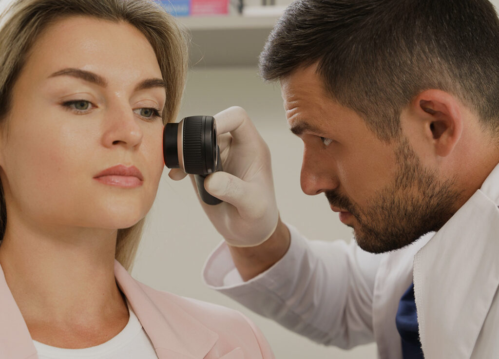
Examine a rare case of basal cell carcinoma arising in the lower lip mucosa, including its unusual presentation and diagnostic challenges.
Examine a rare case of basal cell carcinoma arising in the lower lip mucosa, including its unusual presentation and diagnostic challenges.

Review the diagnosis and management of acral pigmented lesions, including dermoscopic clues that distinguish benign lesions from acral melanoma.

See how an invasive melanoma concealed within tattoo pigment appeared featureless, highlighting the diagnostic risks of examining tattooed skin.

Discover why dermatoscope training belongs in primary care and how short, structured education can improve skin tumour triage and diagnostic performance.

Review melanoma diagnosis and treatment delays across primary care and hospital services, including where collaboration improved timeliness.

Book a personalised MoleMax HD demo to explore digital dermoscopy, lesion monitoring and clinical workflow tools with a product specialist.
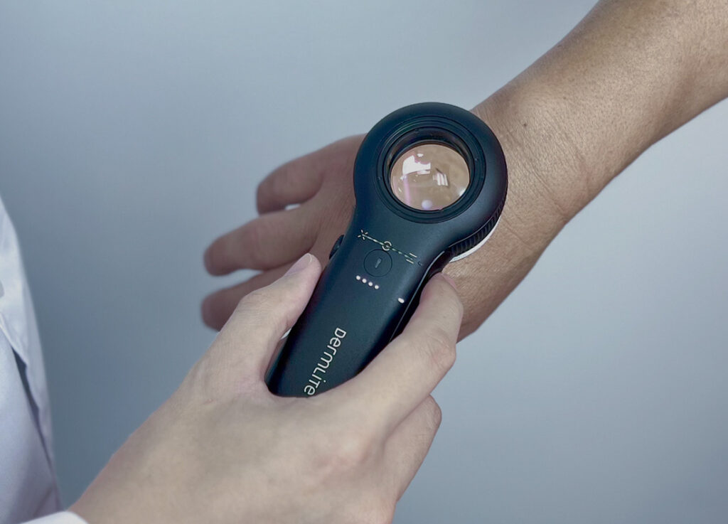
In inflammatory and infectious diseases, the main histopathologic alterations are usually not associated with pigment, but include cellular infiltrations, vascular structures and alterations of the thickness or the anatomy of…

Compare basal cell carcinoma dermoscopic patterns in facial H-zones and non-H-zones, including differences in ulceration and brown globules.

Explore expert recommendations for smarter European e-skin cancer care, addressing delayed diagnosis, unequal access and post-pandemic digital pathways.
Review how cutaneous lichen planus dermoscopy terminology can shift from metaphoric labels to clearer, standardised descriptive language.

See how a short online course improved primary care physicians’ knowledge of important nail diseases, diagnosis and management.

Review International Dermoscopy Society recommendations for selecting patients with multiple nevi for total body photography and digital monitoring.
MoleMax Systems, a division of Macquarie Medical Systems, specialises in the development and production of dermatoscopic hardware and software, including dermatoscopes and the MoleMax Dermoscopy Software. These integrated solutions are designed for early clinical imaging sessions, analysis and comparisons of skin cancer, providing dermatologists with advanced tools for high-resolution imaging and analysis of skin lesions to improve patient outcomes.
Subscribe To The MoleMax Newsletter