Rinni R Patel, Hita H Mehta, Manal D Dave
Abstract
Introduction: Follicular dermatoses are conditions characterized by localization around hair follicles, often presenting as small papular lesions. These dermatoses can be challenging to diagnose with the naked eye, making dermoscopy an important adjunctive diagnostic tool. However, the literature on the dermoscopic features of follicular dermatoses remains limited.
Objectives: To evaluate dermoscopic findings in follicular dermatoses.
Methods: This cross-sectional study was conducted from November 2022 to January 2024 at the dermatology department of a tertiary hospital. Patients were categorized into inflammatory and keratinization follicular dermatosis. A fully developed lesion was dermoscopically examined using a DermLite DL-5 dermoscope. The data were statistically analyzed.
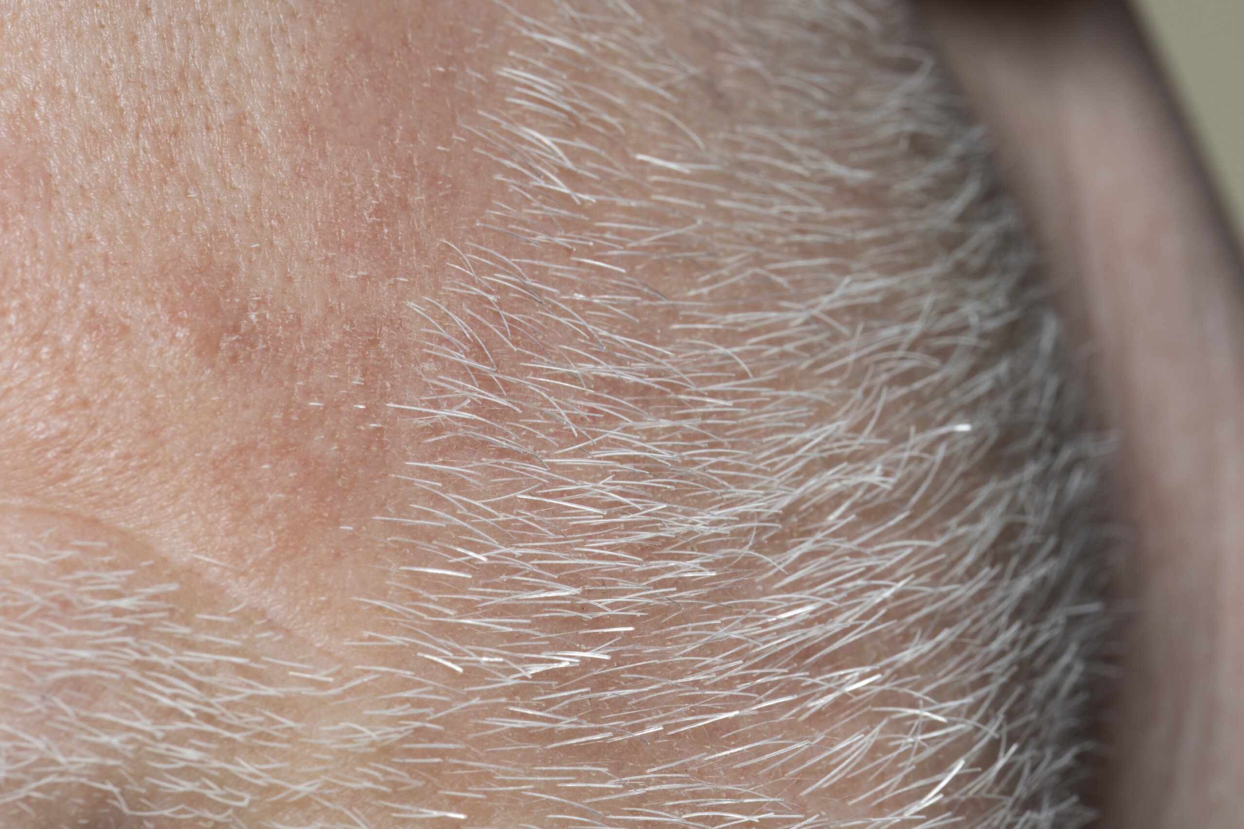
Results: We studied 147 patients. Most follicular dermatoses on dermoscopy showed classical findings such as keratotic plugs, perifollicular white halo, or brown halo. However, they also exhibited characteristic features unique to each dermatoses. Keratosis pilaris exhibited coiled hair (77.41%). Reactive perforating collagenosis presented a central yellowish plug (100%), whitish rim (100%), and peripheral erythematous halo (87.5%). Follicular LP showed reduced follicular ostia (45.45%) and blue-gray globules (40.90%), with newer findings like radial white stria and rosette. Follicular psoriasis displayed regular red dots (100%), while follicular eczema showed red globules (50%) and irregular red dots (16.7%). Acne keloidalis nuchae exhibited radial white streaks (62.5%), perifollicular white globules (50%), V-shaped hair (50%), and radial linear vessels (18.75%). Darier disease showed central hyperpigmented and white keratotic plugs, comedo-like openings, and interfollicular exaggerated pseudo-pigment areas.
Conclusion: Our study highlights that each follicular dermatosis presents specific dermoscopic patterns, supporting dermoscopy as a useful, noninvasive tool for differentiation that can complement histopathology.
To read the full article please click here.