Hair loss is one of the most common reasons patients seek dermatological help, yet it is notoriously difficult to assess objectively by eye. Trichoscopy, dermoscopy of the hair and scalp, has transformed this field by making it possible to examine, measure, and monitor hair with real precision. This guide explains what trichoscopy is, how automated tools such as TrichoScan work, and why objective hair analysis matters in modern practice.
What Is Trichoscopy?
Trichoscopy is the application of dermoscopy to the scalp and hair. Using magnification and controlled illumination, it lets clinicians examine hair shaft thickness, follicular openings, perifollicular skin, and vascular patterns that are invisible to the naked eye. It has become a first-line, non-invasive tool for diagnosing hair and scalp disorders, often reducing the need for a more invasive scalp biopsy.
The technique is valuable because different conditions produce different, recognisable patterns. Androgenetic alopecia, alopecia areata, telogen effluvium, and scarring alopecias each show characteristic trichoscopic features, allowing a more confident diagnosis at the bedside.
Why Objective Hair Measurement Matters
The core problem in managing hair loss is measurement. Patients and clinicians alike struggle to judge, by memory or photographs, whether hair is thinning, stable, or regrowing. Subjective assessment is unreliable and slow to show change. Objective hair density analysis solves this by turning the scalp into measurable numbers hairs per square centimetre, average shaft thickness, and the ratio of growing to resting hairs that can be tracked over time.
How TrichoScan Works
TrichoScan is a trichoscopy software system that combines epiluminescence microscopy with automatic image analysis to measure the biological parameters of hair growth in a single clinical workflow. In practice, a small area of scalp is imaged, and the software automatically counts and characterises the hairs, measuring hair density, hair diameter, and the anagen (growing) to telogen (resting) ratio. Because the analysis is automated, it removes much of the subjectivity and variability of manual counting.
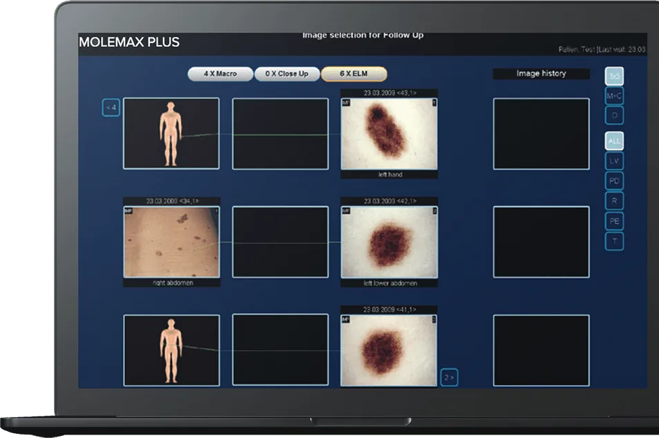
The TrichoScan Software is used as an add-on within the MoleMax imaging ecosystem and is clinically validated with a high operator-agreement rate. That reproducibility is what makes it suitable not just for diagnosis but for objectively monitoring how a patient responds to treatment over months.
Key Parameters Trichoscopy Measures
Hair density. The number of hairs per unit area the headline figure for tracking thinning or regrowth.
Hair shaft diameter. The thickness of individual hairs. Progressive miniaturisation (thinning shafts) is a hallmark of androgenetic alopecia.
Anagen-to-telogen ratio. The proportion of hairs actively growing versus resting. A shift toward telogen signals increased shedding, as seen in telogen effluvium.
Follicular units. The number of hairs per follicular unit, another marker of miniaturisation over time.
Clinical Uses of Trichoscopy and TrichoScan
The most common application is androgenetic alopecia, male and female pattern hair loss, where objective density and diameter measurements confirm the diagnosis and establish a baseline. Trichoscopy is equally useful for distinguishing scarring from non-scarring alopecias, assessing alopecia areata, and evaluating telogen effluvium. Reference resources such as DermNet describe the characteristic trichoscopic patterns for each of these conditions in detail.
Beyond diagnosis, the biggest practical value is treatment monitoring. When a patient starts therapy, repeat TrichoScan measurements at intervals show, in hard numbers, whether the treatment is working information that supports better decisions and keeps patients engaged with their care.
Trichoscopy in a Skin Imaging Clinic
For clinics already performing digital dermoscopy, adding trichoscopy is a natural extension. The same principles of magnified imaging and structured documentation apply, and a system like the MoleMax HD can support both skin and scalp imaging within one workflow. Handheld dermatoscopes from the DermLite range can also be used for quick scalp examination, with some models offering hair-counting contact plates. This makes trichoscopy an efficient way to broaden the services a clinic offers without a wholesale change in equipment.
A Note on Terminology
Patients and referrers search for this capability in many ways, trichoscan test, trichoscan machine, hair analysis, and trichoscopy among them. Using these natural terms in your patient-facing materials helps the people who need the service actually find it, since the underlying demand, as search data shows, is considerable and, at present, largely underserved.
Trichoscopy vs Traditional Methods
Before trichoscopy, clinicians relied on methods that were either crude or invasive. The hair-pull test gives only a rough sense of active shedding. Manual hair counts are tedious and inconsistent. The classic phototrichogram, while informative, is labour-intensive. Scalp biopsy, though sometimes necessary, is invasive and unpopular with patients. Trichoscopy, and automated analysis in particular, offers most of the diagnostic value of these methods non-invasively and in minutes, which is why it has become a first-line approach for hair and scalp assessment.
Common Trichoscopic Patterns by Condition
Part of what makes trichoscopy so powerful is that conditions produce recognisable signatures. Androgenetic alopecia typically shows hair-diameter diversity (a mix of thick and miniaturised hairs), a higher proportion of single-hair follicular units, and peripilar changes, concentrated over the crown. Alopecia areata characteristically shows exclamation-mark hairs, black dots, and yellow dots. Telogen effluvium shows increased empty follicles and regrowing short hairs without the miniaturisation of androgenetic loss. Scarring alopecias show loss of follicular openings, a key warning sign that prompts urgent management.
Recognising these patterns lets a clinician reach a confident diagnosis at the first visit in many cases, and objective measurement then anchors the plan that follows.
Setting Up Trichoscopy in Practice
Introducing trichoscopy does not require rebuilding a clinic. At its simplest, a handheld dermatoscope and a consistent technique are enough to begin recognising patterns. Adding an automated system such as TrichoScan Software brings reproducible measurement, which is what elevates trichoscopy from a diagnostic aid to a monitoring tool. Standardising the imaging site, lighting, and, where required, preparation of the scalp area ensures that measurements taken months apart are genuinely comparable.
Consistency is everything in monitoring. A measurement is only meaningful against a comparable baseline, so establishing a repeatable protocol from the outset is the most important step a clinic can take.
The Patient Experience
Trichoscopy also changes the conversation with patients, much as skin dermoscopy does. Being able to show someone a magnified image of their own scalp, and later a set of numbers demonstrating that a treatment is working, is far more persuasive than reassurance alone. That objective feedback improves adherence to treatment and helps set realistic expectations both of which matter in a field where results take months and patient anxiety is common.
Limitations to Keep in Mind
Trichoscopy is powerful, but it is not infallible. Automated measurement depends on good technique — consistent imaging site, adequate contrast, and proper preparation of the area and poor image quality produces unreliable numbers. Some overlapping conditions can share trichoscopic features, so findings must always be interpreted alongside the clinical history and examination. And while trichoscopy reduces the need for scalp biopsy, it does not eliminate it entirely; scarring alopecias in particular may still require histology. Treating trichoscopy as a decision-support tool rather than a standalone diagnosis keeps its use appropriately grounded.
Combining Trichoscopy with Overall Skin Care
For a clinic, trichoscopy rarely stands alone it complements a broader skin and imaging service. Patients presenting with hair concerns often have other dermatological needs, and a practice already equipped for digital dermoscopy is well placed to offer scalp assessment within the same visit. Integrating hair analysis into an existing imaging workflow spreads the value of the equipment across more of the patient base and positions the clinic as a comprehensive skin-health provider rather than a single-service one. Over time, that breadth is both a clinical and a commercial advantage.
The Takeaway
Trichoscopy has turned hair and scalp assessment from an impression into a measurement. It supports faster, more confident diagnosis of common conditions like androgenetic alopecia, and crucially it lets clinicians prove whether treatment is working through objective, repeatable numbers. For a skin-imaging clinic, adding trichoscopy is a natural, low-friction way to broaden services, deepen patient trust, and meet a clear and currently underserved demand. The technology exists, the search interest is there, and the clinical case is well established; the remaining step is simply putting it to work.
Add Objective Hair Analysis to Your Clinic
The best way to choose is to try the technology in your own clinic. Book a free 15-minute MoleMax demo, and our specialist team will help you match the right dermatoscope or imaging system to your workflow, patient volume, and budget.