Melanoma diagnosed at the earliest stage has a five-year relative survival rate of around 99%; once it has spread to distant sites, that figure falls to roughly 26%. The gap between those two numbers is why clinics invest in dedicated screening equipment rather than relying on the naked eye alone.
This guide is a buying and equipment guide: it compares the categories of skin cancer detection devices on the market, what to evaluate before purchasing, and where the MoleMax range fits. It intentionally does not re-explain how AI lesion analysis works step by step for that mechanism see our in-depth explainer, AI Skin Cancer Detection System: How It Works and Why Clinics Are Adopting It.
What Counts as a Skin Cancer Detection Device
Three equipment families cover most clinical use:
- Handheld dermatoscopes magnify and illuminate a single lesion for immediate visual assessment. They capture nothing automatically unless paired with a camera or phone.
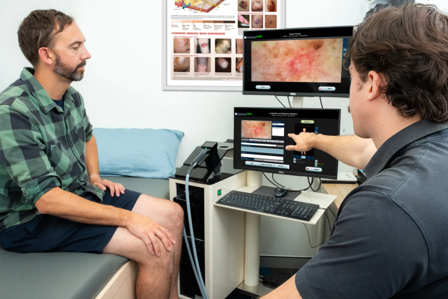
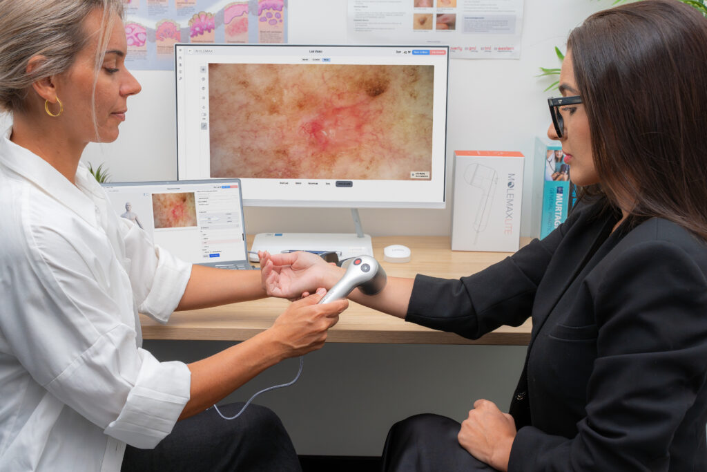
- Digital dermoscopy systems capture, store and track images across visits, turning a one-off look into a longitudinal record.
- Point-of-care adjunctive devices use a non-imaging technology spectroscopy or impedance measurement rather than a photograph, to score a single lesion’s risk at the point of contact.
Most dermatology clinics combine the first two. The third category is newer and sits mainly in primary care; the section below places it in context.
The Device Landscape, by Category

Knowing where a product sits in the wider market helps when a demo or a sales conversation starts comparing systems.
| Category | What it does | Regulatory note | Example products |
| Handheld dermatoscope | Magnifies and illuminates one lesion; no storage on its own | General-purpose optical instrument | DermLite range |
| Digital dermoscopy / total body system | Captures, stores and tracks images across visits; some add AI-assisted scoring | Varies by market and software claims | MoleMax HD, MoleMax HD PRO, FotoFinder, Canfield Scientific |
| Point-of-care adjunctive device | Scores a single lesion using spectroscopy or impedance rather than a stored photograph; used as a rule-out aid, not a diagnosis | FDA-cleared examples exist for primary care use (e.g., DermaSensor, cleared for use by non-specialists; MelaFind and NeviSense were earlier cleared devices in this category) | DermaSensor, MelaFind, NeviSense |
These adjunctive devices are built for a different setting primary care triage of a single concerning lesion rather than the ongoing, whole-body monitoring that a dermatology or skin-cancer clinic typically needs. For a deeper comparison of AI-based melanoma detection systems, accuracy figures and limitations, see our AI Melanoma Detection System for Clinics: A 2026 Guide.
Total Body vs. Single-Lesion Imaging, in Brief
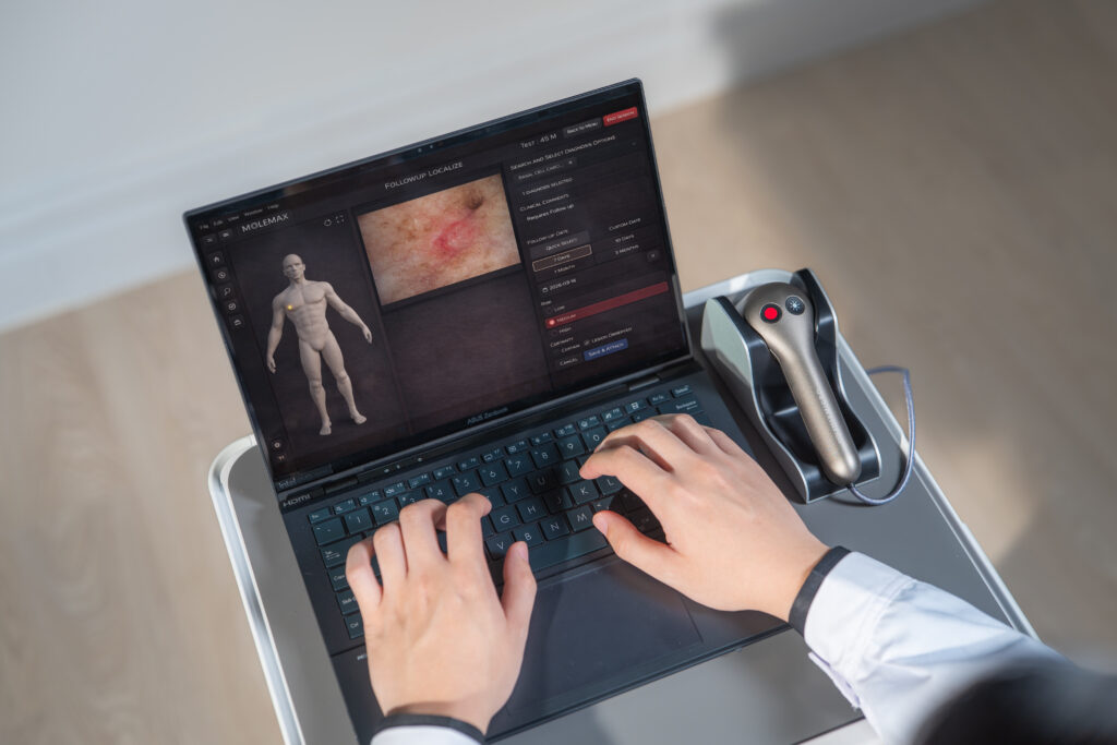
Single-lesion dermoscopy zooms in on one spot; total body photography records the whole skin surface so new or changing lesions stand out at a glance on the next visit. Most well-equipped clinics run both, linking an overview image to a detailed dermoscopic capture of anything worth a closer look.
For the full mechanics of mole mapping, total body photography, dermoscopy and change tracking together, see Mole Mapping Technology: What It Is, How It Works, and Why Clinics Are Adopting It.
Where the MoleMax Range Fits
| Product | Category | Best fit |
| DermLite dermatoscopes | Handheld dermatoscope | First purchase for a clinic new to dermoscopy, or occasional single-lesion checks |
| MoleMax Lite | Digital dermoscopy, entry-level | Clinics wanting digital capture without a full total-body setup, with room to upgrade later |
| MoleMax HD | Digital dermoscopy, full system | Clinics screening regularly, needing lesion scoring and structured reporting |
| MoleMax HD PRO | Digital dermoscopy with motorised total body mapping | High-volume skin-cancer clinics running full-body baseline and follow-up imaging |
| TrichoScan | Specialised imaging | Scalp and hair density assessment — outside lesion detection, but built on the same image-analysis approach |
A practice can start with a MoleMax Lite and move up to a MoleMax HD or HD PRO as patient volume and total-body-mapping needs grow, without discarding the initial investment.
What to Evaluate Before Buying
| Factor | Questions to ask |
| Image quality | Resolution, polarised vs. non-polarised capture, consistency of lighting across sessions |
| Change tracking | Can the software place two visits’ images side by side automatically, or does staff do this manually? |
| Reporting | Does it generate a structured, patient-ready report linked to the patient record? |
| Regulatory status | Is the device/software cleared or registered for its claimed use in your market (e.g., TGA in Australia, FDA in the US)? |
| Total cost of ownership | Upfront hardware cost, software licensing, any recurring subscription fee, and support/training cost |
| Training and support | Onsite or remote training included; typical time for staff to reach confident daily use |
On cost: not every system in this category charges a subscription confirm this specifically, since it materially changes total cost of ownership over a 3–5 year horizon, not just the headline price.
Why This Matters for Outcomes
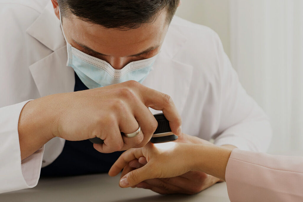
Cancer Australia reports an overall 94% five-year relative survival rate for melanoma diagnosed in 2017–2021. Survival varies sharply by stage at diagnosis: Cancer Council Australia cites close to 100% five-year survival for Stage I melanoma, falling to roughly 26% for Stage IV. The American Academy of Dermatology similarly recommends regular skin self-exams and professional skin checks as central to catching melanoma while it is still thin and localised.
Digital monitoring doesn’t replace clinical judgement, but it directly supports this stage-at-diagnosis effect: a stored image history makes a slow-changing lesion visible months before it would otherwise prompt a visit. For how AI-assisted systems perform against dermatologists of varying experience in realistic settings, including the gap between expert and novice readers, see our clinical-evidence review, Limits of Artificial Intelligence Models for Skin Cancer Diagnosis in Realistic Settings.
Fitting a Device Into the Clinic Day
A typical rollout: capture a baseline full-body image set at a patient’s first skin check, dermoscopically image any lesion of concern, then schedule a follow-up so the same lesions are re-examined and compared automatically. With proper training, most clinics integrate digital dermoscopy within a few weeks, and the documentation time saved tends to offset the learning curve quickly.
Being able to show a patient a magnified image of their own lesion and explain what’s being monitored and why also improves follow-up attendance, which matters as much for outcomes as the imaging itself.
Common Procurement Questions
Do I need a full digital dermoscopy system, or is a dermatoscope enough? For occasional checks, a handheld dermatoscope is usually enough. Clinics screening regularly, or monitoring high-risk patients over time, get the most value from digital capture with change tracking.
Is a subscription required? Not with every system. Some digital dermoscopy platforms, including the MoleMax range, are offered without ongoing subscription fees. Confirm this before comparing headline prices, since it changes the multi-year cost picture considerably.
Can I start small and upgrade later? Yes, within the same product family, a MoleMax Lite deployment can move to a MoleMax HD or HD PRO as volume grows, without re-doing the initial setup.
How long does staff training take? Most clinics report confident daily use within a few weeks of onsite or remote training; exact timing depends on how much the team documents today versus on paper.
Does total body mapping require a dedicated room? The motorised total-body-mapping stand used with systems like the MoleMax HD PRO needs a fixed, consistent-lighting space; a lighter dermoscopy-only setup does not.
See It in Your Own Workflow
Book a free 15-minute MoleMax demo to see high-resolution capture, lesion tracking, side-by-side comparison and structured reporting on real cases, and get your specific procurement questions answered.