Skin cancer is one of the most common cancers globally and one of the most survivable when caught early. Mole mapping technology is changing how dermatologists detect and monitor suspicious skin changes before they become life-threatening. If you are a clinic owner evaluating this system, or a patient trying to understand what the procedure involves, this guide covers everything you need to make an informed decision.
What Is Mole Mapping Technology?
Mole mapping technology is a clinical imaging system that photographs, documents, and digitally stores every mole and skin lesion on a patient’s body. It uses high-resolution cameras combined with specialized software to create a full-body skin record that is compared across multiple visits over time. The goal is not to diagnose cancer in a single session, it is to detect subtle changes in moles that may indicate early melanoma before they become visible to the naked eye.
The technology works by establishing a baseline at the first appointment. Every subsequent visit compares current images against that baseline, allowing the dermatologist to identify new moles, changes in size or shape, or shifts in color that might otherwise go unnoticed during a standard examination.
How Is It Different from a Regular Skin Check?
A regular skin check is a visual examination conducted during a single appointment, typically with a handheld dermatoscope. It captures what the clinician sees today — but nothing is stored, measured, or compared to a previous visit.
Mole mapping creates a permanent, structured digital record. It does not replace the clinical examination; it adds a longitudinal dimension to it. The difference is between a photograph and a video, one captures a moment, the other captures change.
What Does “Total Body Photography” Mean?
Total body photography is the clinical term used interchangeably with full body mole mapping. It refers to the systematic, standardized photography of the entire skin surface — from scalp to feet, using high-resolution imaging equipment.
It is the imaging step within the broader mole mapping process. The full process also includes individual lesion dermoscopy, AI-assisted analysis, and follow-up comparison. Total body photography is what produces the baseline body map that everything else is built on.
How Does Mole Mapping Work? Step-by-Step
Mole mapping follows a structured five-step process. Here is exactly what happens from the moment a patient walks in to the moment they leave with a follow-up plan.
Step 1: Consultation and Skin History
The appointment begins with a clinical interview. The dermatologist or nurse records the patient’s total mole count, any history of atypical moles, personal or family history of melanoma, and cumulative sun exposure. This information builds a risk profile that determines how frequently the patient should be monitored and whether insurance coverage may apply.
This step also sets expectations. Patients are told what the procedure involves, how images will be stored, and what outcomes to expect from the session.
Step 2: Full-Body Image Capture
The patient is guided through a series of standardized poses while a high-resolution camera system photographs every surface of the body. This step typically takes 15 to 30 minutes. Standardized poses are critical — they ensure that images from one visit can be accurately aligned and compared against images from the next.
Body regions covered include the face, scalp, neck, chest, back, abdomen, arms, hands, legs, and feet. No area is skipped, because melanoma can develop anywhere on the skin, including regions patients rarely examine themselves.
Step 3: Dermoscopic Imaging of Individual Lesions
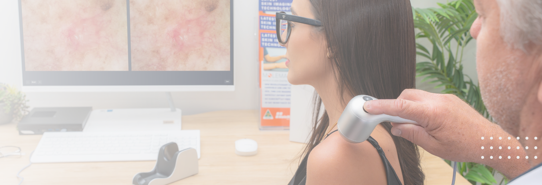
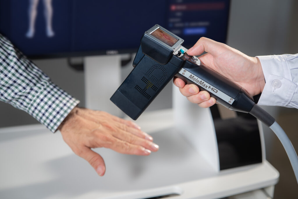
After full-body photography, the dermatologist uses a dermatoscope — a handheld device that magnifies and illuminates the skin’s surface — to capture close-up images of individual moles flagged as atypical or worth monitoring closely.
These dermoscopic images are linked to their precise location on the full-body map, creating a two-layer record: the macro view showing where a mole sits on the body, and the micro view showing its internal structure in detail. This combination is what makes mole mapping significantly more powerful than either technique used alone.
Step 4: AI Analysis and Change Detection
At this stage, the system’s artificial intelligence compares current images against the stored baseline. The AI scans for changes in mole size, shape, color distribution, border irregularity, and texture across every documented lesion.
Crucially, the AI does not diagnose. It flags, scores, and prioritizes. It presents the dermatologist with a ranked list of lesions that have changed most significantly since the last visit, allowing the clinician to focus their attention where it matters most rather than manually reviewing hundreds of stable moles. A 2020 study published in Nature found that a deep learning model outperformed 58 dermatologists in distinguishing malignant melanomas from benign lesions, achieving an AUC of 0.94 — underscoring the clinical value AI brings to this process.
Step 5: Report and Follow-Up Scheduling
The session concludes with a structured report listing all flagged lesions, their body map location, dermoscopic images, and a change summary for returning patients. The dermatologist reviews the report and gives the patient one of three outcomes: discharge with an annual review, a follow-up appointment in six months, or an immediate biopsy referral for a concerning lesion.
The images are stored and become the patient’s permanent skin record — the asset that grows more valuable with every visit.
Who Should Get Mole Mapping?
Mole mapping is suitable for two broad groups: patients with an identified high-risk profile, and general adults who want to establish a proactive baseline before any problems develop.
Which Patient Profiles Benefit Most?
Certain patients have a clinically elevated risk of developing melanoma and are the primary candidates for regular mole mapping:
- Patients with more than 50 moles: The higher the mole count, the greater the statistical likelihood of one undergoing malignant change
- Patients with atypical or dysplastic nevi : Moles with irregular borders, mixed pigmentation, or asymmetric shape that require close longitudinal monitoring
- Patients with a personal history of melanoma: The risk of a second primary melanoma is significantly higher than average
- Patients with a first-degree relative diagnosed with melanoma: Genetic predisposition raises lifetime risk substantially
According to the Melanoma Institute Australia, regular mole mapping in high-risk populations can detect melanoma 80 to 160% earlier than in patients without systematic monitoring.
Is It Only for High-Risk Patients?
No, but the clinical case for it is strongest in high-risk patients. Any adult can request mole mapping as a proactive baseline. The value of a single scan is limited; the real power comes from serial monitoring over two, five, or ten years.
For clinics, this means mole mapping serves two distinct patient groups with different clinical rationales and different conversation approaches. High-risk patients need mole mapping. General adults who want it are making a proactive choice that deserves to be supported, not discouraged.
Types of Mole Mapping Technology Used in Clinics
Not all mole mapping systems are the same. There are three distinct technology tiers in clinical use today and the differences between them have significant implications for diagnostic accuracy, workflow efficiency, and procurement cost.
- Standard Digital Dermoscopy Systems
These are handheld or table-mounted dermoscopes connected to a digital camera and image management software. The clinician selects which moles to photograph based on their own visual assessment during the examination. Images are stored and can be reviewed at follow-up appointments.
This is the entry-level tier. It is suitable for smaller practices with a lower volume of high-risk patients. The core limitation is clinician-dependent selection — lesions the clinician does not flag during the examination will not be imaged, which introduces a potential for missed detection.
- Automated Total Body Photography Systems
These systems use multiple cameras in a standardized array, or a single camera guided through a structured protocol, to photograph the entire body surface systematically in one session. The patient does not need the clinician to decide which moles to capture the system captures everything.
Images are automatically organized into a full-body map and stored for comparison. This tier removes selection bias and ensures comprehensive coverage. It is the standard of care in dedicated melanoma screening clinics and is increasingly being adopted in mid-to-large dermatology practices.
- AI-Integrated Mole Mapping Platforms
The most advanced tier adds a machine learning layer on top of automated imaging. The AI compares current images with stored baseline images, scores each lesion for degree of change, and surfaces the highest-priority cases for dermatologist review.
This tier dramatically reduces the time a dermatologist spends reviewing stable moles — which in a high-volume practice can represent the majority of a follow-up appointment. For clinics managing hundreds of monitored patients, AI-integrated platforms convert mole mapping from a time-intensive process into a scalable clinical program.
- Cost and Insurance Coverage
The cost of mole mapping varies significantly depending on country, clinic type, and the technology tier being used. Insurance coverage exists in some cases but is not guaranteed, and understanding this upfront prevents frustration for both clinics and patients.
How Much Does Mole Mapping Cost?
In the United States, out-of-pocket costs for a full mole mapping session typically range from $150 to $400. This usually includes full-body image capture, dermoscopic imaging of individual lesions, digital storage, and physician analysis. One example pricing structure charges $250 for patients without insurance coverage, covering all four components in a single appointment fee.
The initial baseline appointment is always more expensive than follow-up visits, because it involves establishing the full-body map from scratch. Follow-up appointments focus on comparison and flagging changes, which takes less clinical time and usually costs less.
In Australia and the United Kingdom, costs depend on whether the clinic is public, private, or operating under a national skin cancer screening program. Prices and coverage rules differ significantly across these markets.
Is It Covered by Insurance?
Coverage is inconsistent and should never be assumed. Insurance plans may cover mole mapping for patients with multiple dysplastic nevi, a personal history of melanoma, or a documented family history of melanoma but many insurers do not cover it at all.
Where coverage exists, prior authorization is often required before the appointment. Patients should contact their insurer directly and ask their dermatologist to provide written documentation of medical necessity before booking. It is also important to note that mole mapping is not classified as preventive care under most insurance plans, which means it does not fall under free preventive benefit provisions even for patients with comprehensive coverage.
Why Are Clinics Investing in This Technology?
Clinics adopting mole mapping technology are responding to three converging drivers: demonstrable clinical benefits, rising patient demand for proactive skin care, and a rapidly growing global market that rewards early adoption.
Clinical Benefits for the Practice
The most immediate clinical benefit is a reduction in unnecessary biopsies. When a dermatologist has two years of longitudinal imaging showing a mole has been completely stable, the case for biopsy weakens significantly. Research has shown that baseline mole mapping combined with AI analysis reduces unnecessary biopsies while simultaneously detecting melanoma at an earlier, more treatable stage.
For the practice, fewer unnecessary biopsies means lower pathology costs, less patient anxiety, and more efficient appointment time. A dermatologist who can review an AI-prioritized list of changed lesions rather than manually examining every documented mole in a patient’s record spends their clinical time where it genuinely matters.
Market Size and Growth
The global AI dermatology mole mapping market reached USD 1.47 billion in 2024 and is projected to grow at a compound annual growth rate of 19.2% through 2033, reaching USD 6.25 billion. This growth is driven by rising global skin cancer incidence, rapid advances in AI imaging accuracy, and increasing clinical demand for systematic early detection protocols.
Clinics investing in mole mapping technology now are entering a market that is still in its institutional adoption phase — not saturation. The practices that build mole mapping programs today will hold a significant patient acquisition and retention advantage as demand accelerates over the next decade.
See how MoleMax fits into your clinic’s diagnostic workflow: book a 15-minute product demo today.
Frequently Asked Questions
How Often Should Mole Mapping Be Repeated?
Most dermatologists recommend annual mole mapping for high-risk patients. Patients with rapidly changing lesions or a prior melanoma diagnosis may be scheduled every six months. The interval is always determined by the individual’s risk profile — there is no universal fixed schedule.
Can Mole Mapping Detect Melanoma?
Mole mapping does not diagnose melanoma. Only a biopsy followed by histopathological laboratory analysis can confirm a diagnosis. Mole mapping detects changes in lesions that may warrant a biopsy. Its clinical value lies in identifying suspicious changes at the earliest possible stage, when melanoma is most treatable and outcomes are best.
What Is the Difference Between Mole Mapping and Dermoscopy?
Dermoscopy is a technique using a handheld magnification device to examine individual moles in close detail during a single appointment. Mole mapping is a broader clinical system that uses dermoscopy as one component alongside full-body photography, digital storage, AI analysis, and longitudinal comparison across multiple visits. Dermoscopy examines one mole today; mole mapping tracks all moles over years.
Is Mole Mapping Covered by Insurance?
Coverage depends entirely on the patient’s risk profile and their specific insurance plan. Patients with a documented history of atypical moles, personal melanoma diagnosis, or first-degree relative with melanoma are more likely to qualify for partial or full coverage. Prior authorization is often required. Mole mapping is not classified as preventive care under most plans, so standard preventive benefit provisions do not apply. Always verify coverage with your insurer before the appointment.