Melanoma is responsible for the majority of skin cancer deaths worldwide, yet it is one of the most survivable cancers when detected at an early stage. The problem has always been consistency. Visual diagnosis varies from clinician to clinician, and in under-resourced or primary care settings, that variability costs lives. An AI melanoma detection system for clinics addresses this directly by adding a standardized, data-driven layer to every skin assessment. This guide covers what these systems are, how accurate they are, which ones clinics are using, and why adoption is accelerating in 2026.
What Is an AI Melanoma Detection System?

An AI melanoma detection system is a clinical tool that analyses dermoscopic or photographic images of skin lesions using machine learning algorithms to return a malignancy risk classification. It is trained on large validated datasets such as the ISIC archive and HAM10000, which together contain hundreds of thousands of confirmed skin lesion cases. The system gives the clinician a structured second opinion in seconds. It does not diagnose, and it does not override clinical judgment.
These systems are increasingly embedded into existing AI skin cancer detection software and mole mapping platforms that dermatology clinics already use, meaning adoption does not always require purchasing entirely new infrastructure.
How Does It Differ from a Standard Dermatoscope?
A standard dermatoscope magnifies and illuminates a lesion so the clinician can examine it visually. It produces no output beyond the image itself. An AI melanoma detection system adds an algorithmic layer that processes the image and returns a risk score based on pattern recognition across thousands of confirmed cases. One is a lens. The other is a lens with a trained analytical engine attached.
How Does It Work?
The clinical workflow follows four steps. The clinician captures a high-resolution image of the suspicious lesion using a connected dermatoscope or imaging device. The image is processed by a convolutional neural network trained on datasets like ISIC and HAM10000. The algorithm returns a risk classification, typically low, moderate, or high suspicion for malignancy. The clinician then uses that output alongside their own assessment and the patient’s clinical history to decide the next step: monitor, refer, or biopsy.
The AI handles pattern recognition at speed and scale. The clinician handles context and final judgment.
How Accurate Are AI Melanoma Detection Systems?
The accuracy data published in 2025 makes a strong case for clinical adoption. According to a systematic review published in PMC, modern AI systems achieve pooled sensitivity of 86.3% and specificity of 78.4%, compared to generalist clinicians who achieve sensitivity of just 64.6%. That gap is not marginal. It means a GP using an AI system catches melanoma at a rate far closer to a specialist dermatologist than to an unaided primary care physician.
At the device level, results are even stronger. According to DermaSensor’s FDA pivotal study, conducted across 22 centers and led by the Mayo Clinic, the device achieved 96% sensitivity across all skin cancers identified in over 1,000 patients. In a companion study, missed skin cancers were cut in half, dropping from 18% to 9%, when primary care physicians used the AI device alongside their clinical assessment.
Current Limitations Clinics Should Understand
AI melanoma detection is clinically validated but not without constraints. Three limitations matter most for clinics evaluating these systems.
- Most models have been trained predominantly on images from lighter skin tones, reducing diagnostic reliability across diverse patient populations
- Real-world specificity is consistently lower than published trial results, meaning more false positives in practice than in controlled studies
- Image quality directly affects output accuracy, and poor lighting or incorrect device positioning can produce unreliable scores regardless of algorithm strength
Understanding these limitations does not weaken the case for adoption. It makes informed system selection and proper staff training essential before go-live.
Which AI Melanoma Detection Systems Are Clinics Using in 2026?
Clinics have three main categories of system to choose from, each suited to a different clinical setting and patient volume.
Point-of-Care Devices
DermaSensor is the most validated option in this category. It is the first FDA-cleared AI-powered device for melanoma, basal cell carcinoma, and squamous cell carcinoma, cleared for use by primary care physicians as well as dermatologists. The device uses elastic scattering spectroscopy combined with machine learning to analyse lesion tissue in under 30 seconds without requiring a dermoscopic image. It is designed for GPs and frontline clinicians who need a fast, reliable triage tool at the point of care.
AI-Integrated Dermoscopy Platforms
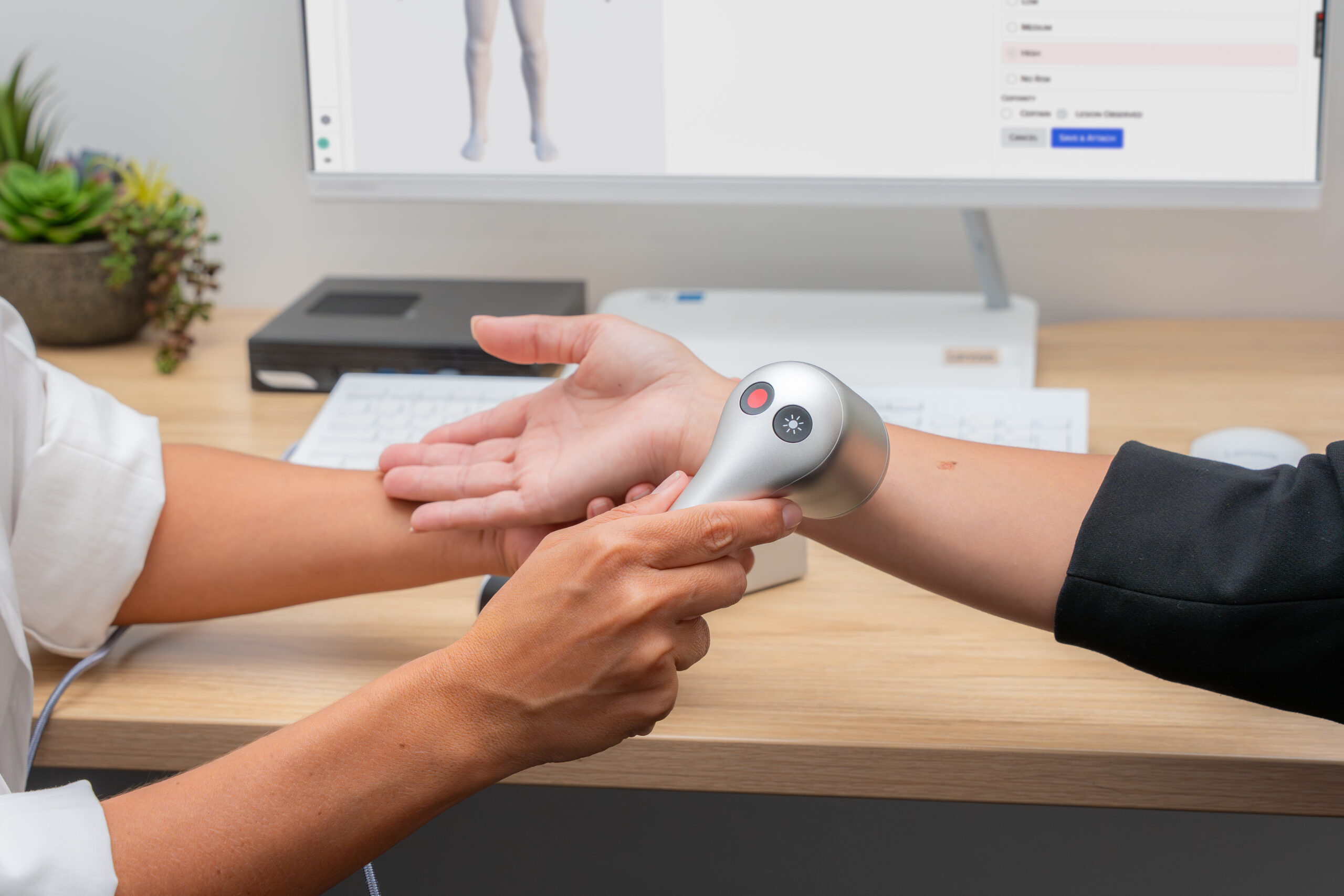
Platforms such as FotoFinder and MoleMax combine high-resolution dermoscopic imaging with an embedded AI layer that compares current lesion images against the patient’s stored historical baseline. This longitudinal comparison is a fundamentally different form of analysis from single-scan risk scoring. It detects subtle changes over time that would be invisible in a one-visit assessment. These platforms are best suited to dermatology clinics and melanoma screening centers running structured mole mapping programs.
App-Based and Consumer Tools
A growing number of AI melanoma detection apps offer image upload and instant analysis directly to patients. These are consumer-grade tools, not clinical-grade systems. They are not FDA-cleared for diagnostic use. Clinics can direct patients toward these tools for self-monitoring between appointments but should communicate clearly that a consumer app result is not a clinical finding and does not replace a professional skin assessment.
How Do Clinics Integrate AI Detection Into Their Workflow?
Integration is simpler than most clinic owners expect. AI melanoma detection fits into three existing touchpoints without requiring a rebuild of clinical processes.
During the appointment, the clinician captures the lesion image and receives a risk score in seconds. The consultation continues normally, with the AI output becoming one input among several rather than a disruptive additional step.
For follow-up monitoring, AI-integrated platforms like MoleMax review stored images between appointments and flag lesions that have changed since the last visit. The dermatologist reviews a prioritized list at the next appointment rather than manually comparing every documented mole. This makes high-volume mole mapping programs clinically manageable at scale.
For GP triage, point-of-care devices help primary care physicians make faster, more confident referral decisions. Instead of referring every uncertain lesion to dermatology and straining specialist capacity, GPs use the AI output to distinguish high-priority referrals from lesions appropriate for watchful waiting.
Why Are Clinics Investing in This Technology?
Three outcomes are driving adoption across dermatology and primary care settings in 2026.
Clinical results improve measurably. Missed melanoma dropped from 29.8% to 20.9% when clinicians used AI assistance in published studies, according to DermaSensor’s clinical utility data. For a clinic running 500 skin cancer consultations per year, that improvement translates directly into earlier diagnoses and better patient outcomes.
The market is growing fast and early movers have an advantage. The global AI dermatology market reached USD 1.47 billion in 2024 and is projected to grow at a compound annual growth rate of 19.2% through 2033, according to DataIntelo’s 2025 market report. Clinics building AI-assisted detection programs now are entering a market still in early institutional adoption, not saturation.
Patient expectations are shifting. Patients are actively seeking clinics that offer AI-assisted skin checks, and in competitive urban markets, early adopters are already differentiating on this capability.
Frequently Asked Questions
Is AI melanoma detection FDA approved?
DermaSensor is FDA-cleared for clinical use at the point of care for all three common skin cancers. Not all AI skin analysis tools carry this clearance. Always verify the regulatory status of any system before clinical adoption.
Can AI detect melanoma from a smartphone photo?
Consumer apps can analyse a smartphone image and return a risk indication, but this is not a clinical diagnosis. Systems used in clinics operate on calibrated dermoscopic images under standardized conditions. The accuracy gap between a clinical AI system and a consumer app is significant.
What is the best AI melanoma detection system for a dermatology clinic?
For high-volume dermatology clinics, an AI-integrated platform like MoleMax that supports longitudinal lesion tracking is the strongest clinical fit. For GPs adding skin cancer screening to their practice, DermaSensor is the most clinically validated point-of-care option available in 2026.
Is there a free AI melanoma detection tool?
Free consumer tools exist for personal monitoring between clinic visits. They are not designed or cleared for clinical diagnostic use. Clinics can recommend them to patients for self-checking but should set clear expectations about their limitations.
AI melanoma detection for clinics is no longer experimental. The clinical evidence is published, the leading devices are FDA-cleared, and workflow integration is proven across primary care and specialist settings. The clinics investing in these systems now are building a diagnostic capability that will define the standard of skin cancer care within the next five years.
See how MoleMax fits your clinic’s diagnostic workflow. Book a 15-minute demo today.