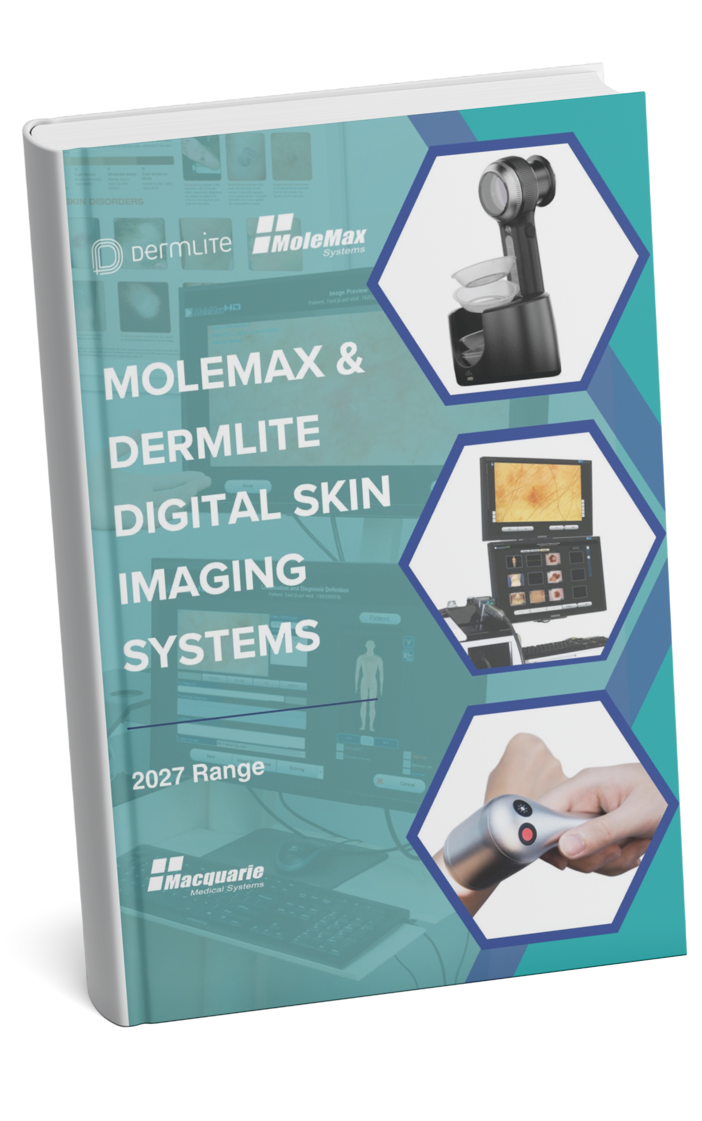
Dermoscopy and digital dermoscopy are closely related but fundamentally different tools in modern skin examination. Both are used to visualise subsurface skin structures not visible to the naked eye, yet they differ significantly in how images are captured, stored, reviewed and used over time.
Understanding these differences is essential for clinics focused on accurate documentation, follow-up and consistent clinical workflow rather than single-visit assessment alone.
What Is Dermoscopy?
Dermoscopy is an optical examination technique using a handheld dermatoscope that provides magnification and specialised lighting. It allows clinicians to assess patterns, colours, and structures within a pigmented skin lesion during a live consultation.
The assessment is immediate and relies heavily on clinician experience and visual interpretation. Findings are typically recorded as written notes, diagrams, or memory-based descriptions.
When Traditional Dermoscopy Is Sufficient
- One-off lesion assessments
- Low-risk patients with limited follow-up needs
- Situations where image storage is not required
- Clinics relying primarily on clinician recall
Dermoscopy remains essential for recognising structures associated with Malignant Through Dermoscopy, particularly when rapid decision-making is required.
What Is Digital Dermoscopy?
Digital dermoscopy builds on traditional dermoscopy by adding image capture, storage and retrieval. Instead of relying only on live optical viewing, images are saved and linked directly to the patient record.
This allows clinicians to:
- Compare lesions across time
- Review images before or after appointments
- Share visual findings during patient discussions
- Support structured documentation and reporting
Digital dermoscopy shifts skin checks from memory-based assessment to evidence-based longitudinal monitoring.
Dermoscopy vs Digital Dermoscopy: Core Differences
Image Capture and Storage
- Dermoscopy: Live optical view only
- Digital dermoscopy: High-quality images saved with patient records
Documentation
- Dermoscopy: Written notes and diagrams
- Digital dermoscopy: Image-linked documentation, measurements, and annotations
Follow-Up
- Dermoscopy: Relies on recall and written descriptions
- Digital dermoscopy: Enables side-by-side image comparison over time
Workflow Consistency
- Dermoscopy: Varies between clinicians
- Digital dermoscopy: Standardised capture and reporting protocols
Clinical Workflow Differences That Matter
In daily practice, the difference is not diagnostic intent but operational reliability. Digital dermoscopy reduces rework caused by missing images, unclear lesion localisation, or incomplete notes. This is particularly relevant when monitoring lesions such as Dysplastic Naevus dermoscopy, where subtle changes over time are clinically meaningful but difficult to recall accurately without images.
Follow-Up and Monitoring Over Time
The true value of digital dermoscopy appears during follow-up. Sequential imaging allows clinicians to confirm stability or detect change without relying on memory. This is critical for patients with multiple lesions or increased skin cancer risk.
Digital systems support:
- Sequential lesion review
- Reduced unnecessary excisions
- Better confidence in “watch and wait” decisions
Follow-up protocols are also commonly discussed alongside preventative strategies, including patient education topics such as Nicotinamide for skin cancer, where ongoing monitoring remains essential even with risk-reduction measures.
Patient Communication and Understanding
Digital dermoscopy improves transparency. Patients can see exactly what the clinician is reviewing, making explanations clearer and improving confidence in management plans.
Visual review is particularly helpful when explaining why a lesion is benign, changing or requires monitoring.
This is especially useful when discussing complex or atypical pigmented skin lesion patterns that are difficult to describe verbally.
Common Misconceptions About Digital Dermoscopy
- It does not replace clinical judgement
- It does not diagnose cancer on its own
- It is not limited to specialist clinics
Digital dermoscopy is a documentation and follow-up tool. Its value lies in consistency, traceability and workflow support rather than standalone diagnostic claims.
How Clinics Decide When to Move Beyond Handheld Dermoscopy
Clinics typically consider digital dermoscopy when:
- Patient volume increases
- Follow-up intervals become routine
- Multiple clinicians need consistent records
- Image-based referrals are required
The transition is usually driven by workflow needs rather than technology preference.
Summary: Dermoscopy vs Digital Dermoscopy
Dermoscopy provides immediate visual assessment. Digital dermoscopy adds documentation, comparison and continuity.
Both tools are clinically valuable, but they serve different roles. Dermoscopy supports moment-in-time evaluation, while digital dermoscopy supports longitudinal care, structured follow-up and consistent communication.
Frequently Asked Questions (FAQs)
Is digital dermoscopy more accurate than dermoscopy?
Digital dermoscopy does not change the optical principles of dermoscopy. Its advantage lies in documentation and comparison, not inherent diagnostic accuracy.
Do all clinics need digital dermoscopy?
No. Clinics with low follow-up volume or predominantly one-off assessments may rely effectively on handheld dermoscopy alone.
Can digital dermoscopy replace biopsies?
No. Digital dermoscopy supports monitoring and decision-making but does not replace histopathological confirmation when clinically indicated.