Skin cancer is one of the most common cancers globally, with melanoma alone responsible for the majority of skin cancer-related deaths worldwide. Early and accurate detection remains the most effective strategy for improving patient outcomes, and the quality of clinical skin examination equipment plays a direct role in that process. For dermatologists, general practitioners, skin cancer clinics, and dermatology nurses, having a reliable, high-performance examination device is not optional, and it is a clinical necessity.
The DermLite Lumio is a portable, cross-polarised skin examination scope designed to meet the daily demands of modern dermatological practice. With a large 75 mm lens, 40 bright white LEDs, and cross-polarisation technology, it delivers clear, consistent visualisation of skin lesions and subsurface structures across a wide range of clinical applications. This article provides a comprehensive overview of the DermLite Lumio, its features, clinical applications, comparative advantages, and why it is a trusted choice for healthcare professionals supported by MoleMax Systems.
What Is the DermLite Lumio?
The DermLite Lumio is a handheld skin examination scope built for dermatologists, dermatology nurses, general practitioners, and skin cancer clinicians who require consistent, high-quality visualisation during both routine and specialised skin assessments.
Unlike standard handheld dermatoscopes designed primarily for individual lesion assessment, the DermLite Lumio is built around a large 75 mm aspheric lens with 2x magnification. This wide-field design provides a significantly broader viewing area, allowing clinicians to assess larger skin regions in a single examination pass. This is particularly valuable in high-volume screening environments where speed and thoroughness must be balanced.
The device incorporates 40 bright white LEDs arranged to provide even, shadow-free illumination across the entire examination field. Combined with cross-polarisation technology, this eliminates surface reflection and glare, enabling detailed visualisation of subsurface skin structures without the need for immersion fluid or physical contact with the skin surface.
The DermLite Lumio is battery-powered, lightweight, and compact, making it well suited for fixed clinic use, ward rounds, domiciliary visits, and mobile practitioners working across multiple care settings. Its design prioritises ease of use without compromising clinical performance.
Key Features of the DermLite Lumio
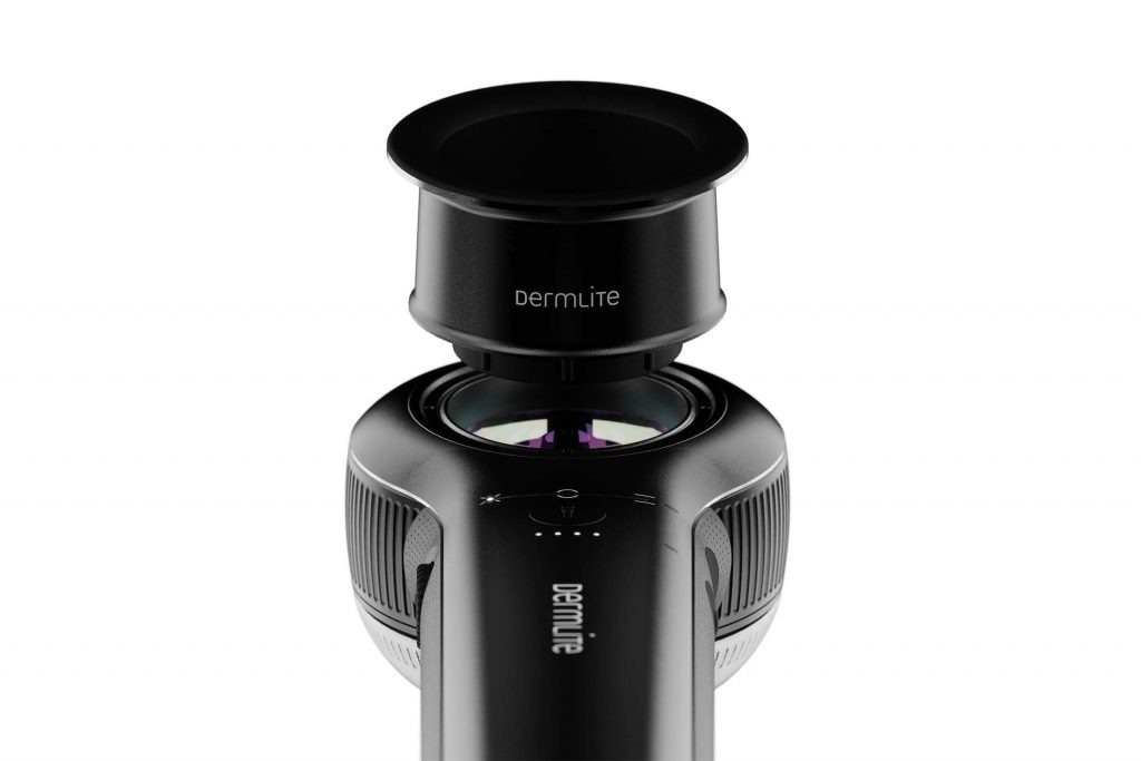
Cross-Polarised Illumination: Cross-polarisation is the defining technical feature of the DermLite Lumio. By filtering light at perpendicular angles, the device suppresses surface reflection and allows clinicians to examine subsurface structures such as pigment networks, vascular patterns, regression structures, and follicular openings with clarity that is impossible under standard white light. This eliminates the need for immersion gel or fluid contact, improving examination hygiene and patient comfort.
Large 75 mm Aspheric Lens: The 75 mm wide-field lens is considerably larger than the typical 20-30 mm viewing area of conventional handheld dermatoscopes. This broader field of view reduces the number of repositioning steps required during each examination, supporting faster assessments and more systematic coverage of the skin surface. The aspheric design minimises optical distortion at the edges of the viewing field, maintaining image quality across the full examination area.
40 Bright White LEDs: Forty LEDs are arranged to deliver even, consistent illumination without hot spots, shadows, or colour variation. Uniform illumination is critical for accurate colour assessment of pigmented lesions and for reliable identification of vascular structures. The LED array is designed for longevity, maintaining consistent light output across thousands of examination cycles.
No Immersion Fluid Required: Cross-polarisation technology removes the requirement for immersion gel or fluid contact during examination. This simplifies the examination process, reduces consumable costs, and improves the patient experience by eliminating a step that many patients find uncomfortable.
Battery-Powered Portability: The DermLite Lumio operates on batteries, removing dependence on power outlets and making it genuinely portable across different clinical environments. Battery life is designed to support a full clinical day of examinations without interruption.
Ergonomic Design: The balanced, lightweight body reduces hand and wrist fatigue during extended examination sessions. The grip and button placement are designed to support one-handed operation, allowing the clinician to maintain patient positioning with the other hand throughout the examination.
Clinical Applications of the DermLite Lumio
The DermLite Lumio is designed as a versatile clinical tool that supports a broad range of dermatological examination needs rather than a single-purpose device.
Pigmented Lesion Assessment: Cross-polarised illumination enables clear visualisation of pigment distribution, border regularity, and internal structural features of pigmented lesions including melanocytic naevi, seborrhoeic keratoses, and suspected melanoma. The ability to assess pigment network architecture and atypical vascular patterns under polarised light supports more informed clinical decision-making about which lesions require further investigation or biopsy.
Skin Cancer Screening Support: The DermLite Lumio supports systematic skin cancer screening workflows where rapid examination of multiple lesions across large body surface areas is required. The wide-field lens reduces examination time per patient, while polarised illumination improves the detection of subtle features that may be missed under naked-eye examination. When used alongside integrated digital imaging platforms such as those offered by MoleMax Systems, the DermLite Lumio contributes to a comprehensive early detection and monitoring workflow.
Hair Follicle and Scalp Examination: The consistent, wide-field illumination of the DermLite Lumio makes it effective for examining hair follicle density, follicular openings, perifollicular scaling, and scalp vascularity. It supports assessment of conditions including androgenetic alopecia, alopecia areata, folliculitis, and seborrhoeic dermatitis.
Vascular Structure Visualisation: Cross-polarised LED illumination enhances visualisation of subsurface vascular patterns including telangiectasias, arborising vessels, dotted vessels, and milky-red areas. These vascular features are diagnostically significant across a range of conditions including basal cell carcinoma, rosacea, and port wine stains.
Varicose Vein and Superficial Vascular Assessment: The large lens and even LED illumination support assessment of varicose veins, reticular veins, and spider telangiectasias, particularly useful in phlebology clinics and vascular dermatology settings where systematic mapping of superficial vessels is required.
Inflammatory and General Dermatological Conditions: Conditions including psoriasis, eczema, lichen planus, pityriasis rosea, and other inflammatory dermatoses benefit from enhanced illumination and magnification during clinical assessment. The wide-field view allows clinicians to assess the distribution and character of lesions across larger skin areas in a single examination step.
Wound and Ulcer Assessment: The DermLite Lumio’s consistent illumination supports clinical assessment of wound margins, granulation tissue, and early signs of infection in chronic wound management settings.
Benefits of Using DermLite Lumio in Clinical Practice
Faster and More Systematic Assessments: The large 75 mm field of view reduces repositioning steps and supports faster, more systematic skin examination. In busy skin cancer clinics and general practice settings, this translates to improved patient throughput without compromising examination quality.
Enhanced Diagnostic Visibility: Cross-polarised illumination consistently reveals subsurface detail that is invisible under standard white light or naked-eye examination. Clinicians benefit from greater confidence in identifying lesion features that influence clinical decision-making, including decisions about biopsy, excision, or monitoring.
Improved Patient Comfort and Experience: Examinations are faster, and the absence of immersion fluid makes them more comfortable. Patients benefit from shorter examination times and a cleaner, less invasive examination process. This is particularly valuable in paediatric dermatology and for patients with sensitive skin conditions.
Reliable Performance Across High-Volume Workloads: The DermLite Lumio is designed for repeated daily use. Its LED array maintains consistent light output, its battery supports full-day operation, and its ergonomic design reduces the physical strain associated with high-volume examination workloads.
Accessible for All Clinical Levels: The DermLite Lumio’s straightforward operation makes it effective for dermatology nurses, general practitioners, registrars, and other team members without specialised dermoscopy training. This broadens its utility across multidisciplinary clinical teams and supports consistent examination quality at all levels of clinical seniority.
Cost-Effective Clinical Investment: Compared with full digital imaging systems, the DermLite Lumio represents a cost-effective entry point into enhanced skin examination capability. For clinics building their dermatology equipment portfolio, it provides immediate diagnostic value while integrating naturally with more advanced systems as the practice grows.