Have you ever looked at a mole and wondered if it could mean something more? It’s important to pay attention to the tiny spots on your skin because that tiny dot may turn into cancer. Mole mapping is a medical procedure that uses high-resolution photography and digital dermoscopic imaging to create a detailed, visual record of all your moles.
In this blog, you’ll learn exactly what mole mapping is, how it works, and which mole changes should be checked early to prevent severe skin cancer risks.
Mole Mapping
Mole mapping is a medical procedure that uses high-resolution photography and digital dermoscopic imaging to create a detailed record of all the moles on your body.
Dermatologists, general practitioners, and skin health practitioners use these images during follow-up visits to compare changes over time, helping them detect suspicious growths or early signs of skin cancer before they become serious. Not every mole turns cancerous, but keeping an eye on them is important because early detection makes treatment much easier and more effective.
What Do the Moles On Your Body Mean?
Moles are harmless skin growths formed by clusters of pigment-producing cells called melanocytes. Most moles are normal, but some can indicate a higher risk of skin cancer, especially if they change in size, shape, or colour. Moles can last more than 50 years. It’s important to see a healthcare provider or dermatologist if you notice any suspicious changes in your skin. They use advanced digital devices to examine moles closely and track their evolution.
What Happens During Mole Mapping?
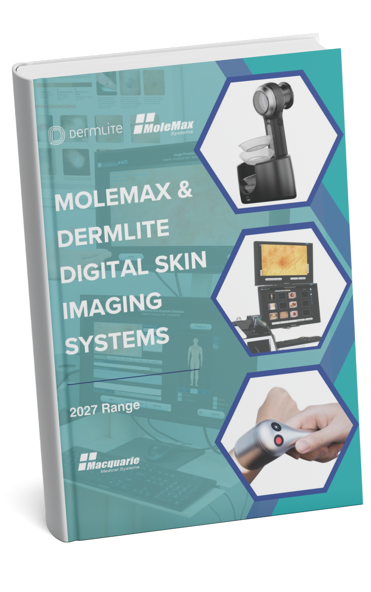
During mole mapping, a medical practitioner or skin health practitioner takes high-resolution photos of your entire body and closely examines each mole using a dermatoscope. They document and highlight any moles that look unusual or have the potential to turn into melanoma or other types of skin cancer. These images are then stored and compared over time to spot even the smallest changes early. Dermatologists and medical practitioners use advanced digital systems like MoleMax, which provide clear images to support precise diagnosis and early skin cancer detection.
How Mole Mapping Works with Advanced Systems Like MoleMax?
MoleMax software is an effective, painless system that uses high-resolution photography and dermoscopic imaging to document, monitor and track moles. Detecting changes early helps protect you from potential skin cancer.
Here’s the Mole Mapping Process with MoleMax:
1. Total Body Photography (TBP)
MoleMax captures detailed, standardised photos of your entire body to record every mole in its exact location. This creates a baseline for future comparisons and ensures that no mole goes unnoticed.
2. Close-Up Dermatoscopic Imaging
Molemax HD pro device used to get a close-up image:
- Examine structure, colour and patterns in detail (ABCDE) method.
- Detect subtle changes that are invisible to the naked eye.
- Track mole evolution over time for early skin cancer detection.
3. Digital Storage and Tracking
All images are securely stored in the MoleMax System. During follow-ups, your medical professional can compare new images with previous ones, detecting even subtle changes over time.
4. Early Detection Alerts
The MoleMax System highlights moles that show irregularities or changes, which allows the medical professional to prioritise examination and intervene early if necessary, reducing the risk of skin cancer.
5. Patient Awareness and Education
Patients can view their mole maps, understand their skin health and learn which moles to monitor, encouraging regular self-checks between appointments.